Login
Sign Up
Member Area
Login
Sign Up
English
Afrikaans formal
Afrikaans informal
Arabic
Azerbaijan
Brazilian
Bulgarian
Catalan
Chinese simplified
Chinese traditional
Croatian
Czech
Danish
Dutch
English
Estonian
Finnish
French
Georgian
German
Greek
Hebrew
Hungarian
Icelandic
Indonesian
Italian
Japanese
Latvian
Lithuanian
Malaysian
Norwegian
Persian
Polish
Portuguese
Romanian
Russian
Serbian
Slovakian
Slovenian
Spanish
Swedish
Thai
Turkish
Ukrainian
Vietnamese
Toggle navigation
VideotonStock
Your Shopping Cart is empty.
Files
Stock
Photo
Video
Special offer
Collections
Packages
Lightboxes
Licenses
Royalty free
Rights managed
Creative
Editorial
Items
Most downloaded
Featured
Most popular
New
Free
Categories
Browse categories
Site info
Site info
About
Support
Privacy Policy
Privacy Policy
FAQ
Contact Us
Contact Us
Terms and Conditions
Buyer Agreement
Seller Agreement
Customers
Users
Credits
Photographers
Boby Richards
0
Boby Richards
0
Videoton1
286
Search
Keywords:
Creator ID:
Type:
Photo
Illustration
Vector
Video
Template
3d
Categories:
Animals
Buildings and Architecture
Business
Drinks
The Environment
States of Mind
Food
Graphic Resources
Hobbies and Leisure
Industry
Landscapes
Lifestyle
People
Plants and Flowers
Culture and Religion
Science
Social Issues
Sports
Technology
Transport
Travel
News
Entertainment
Sport News
Languages:
Brazilan Portuguese - Brazil
English - Canada
French - Canada
Spanish - Mexico
English - United States
English - Belgium
French - Belgium
Dutch - Belgium
Czech - Czech Republic
English - Cyprus
Danish - Danmark
German - Germany
English - Estonia
Spanish - Spain
French - France
French - Marocco
English - Greece
English - Ireland
Italian - Italia
English - Latvia
English - Lithuania
German - Luxembourg
English - Luxembourg
French - Luxembourg
English - Hungary
English - Malta
Dutch - Nederlands
Bokmal Norwegian - Norway
German - Austria
Polish - Poland
Portuguese - Portugal
English - Romania
German - Switzerland
English - Slovenia
Slovakian - Slovakia
French - Switzerland
English - Finland
Swedish- Sweden
Italian - Switzerland
English - United Kingdom
English - Bulgaria
English - Australia
Japanese - Japan
Korean - South Korea
English - New Zealand
Russian - Russia
English - Ukrain
English - Thailand
Orientation:
All
Horizontal
Vertical
square
Color:
Model property release:
All
Yes
No
Duration Video:
All
Up to 10 seconds
Up to 20 seconds
Longer than 30 seconds
Results
(0)
Site
Getty/iStock
Bigstockphoto
Pixabay
Adobe Stock
Relevance
New
Most popular
Most downloaded
Undiscovered
12
24
36
48
96
Auto paging
Menu
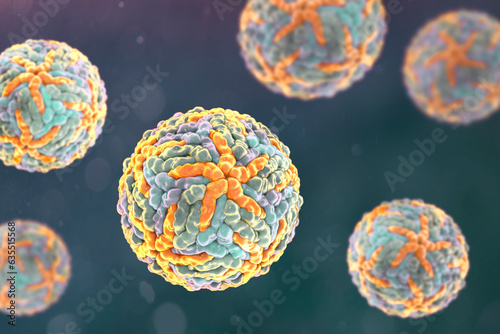
Enteroviruses, 3D illustration showing spherical, non-enveloped RNA viruses responsible for diseases like meningitis, myocarditis, hand-foot-mouth disease.
Mirizzi syndrome, 3D illustration showing compression of the common hepatic duct by an impacted gallstone in the cystic duct, causing obstructive jaundice and inflammation.
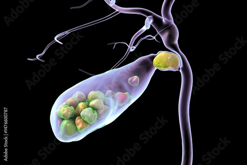
Gallbladder with stones, 3D illustration showing cholelithiasis. Gallstones can block bile flow and cause pain, inflammation, or infection requiring medical or surgical treatment.
Gallbladder with stones, 3D illustration showing cholelithiasis. Gallstones can block bile flow and cause pain, inflammation, or infection requiring medical or surgical treatment.
Anterior cingulate cortex, 3D illustration. Shown in red on a brain mid-section. This region regulates emotion, decision-making, and is implicated in depression and chronic pain.
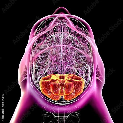
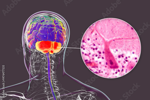
Human brain with highlighted cerebellum, 3D illustration. Cerebellum is responsible for coordination, balance, and fine motor control. Damaged in ataxia and neurodegenerative disorders
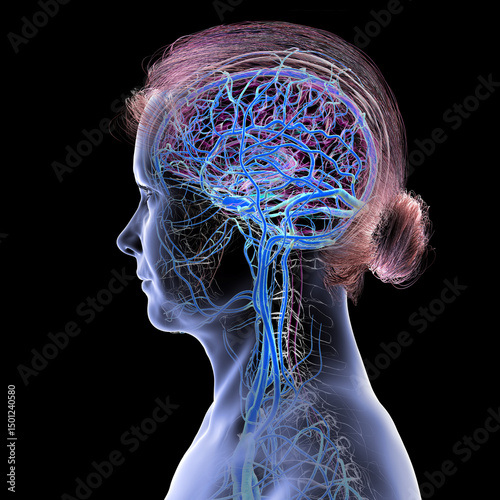
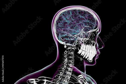
Brain veins, 3D illustration
Clostridium botulinum, 3D illustration. Bacterium producing botulinum toxin, leading to botulism with flaccid paralysis and respiratory failure.
Cerebellum highlighted in colored composite image of magnetic resonance imaging MRI scan of the brain, showing structure involved in balance, coordination, and motor control.
Gallbladder with stones, 3D illustration showing cholelithiasis. Gallstones can block bile flow and cause pain, inflammation, or infection requiring medical or surgical treatment.
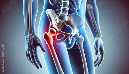
Pain in the hip joint, highlighting conditions such as femur neck fracture, arthritis, and hip dislocation, with emphasis on affected areas. Generative AI illustration.
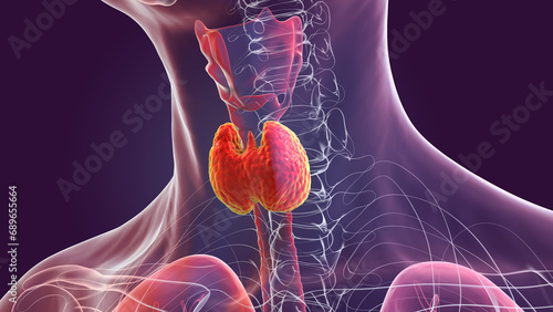
A thyroid gland anatomy, 3D illustration
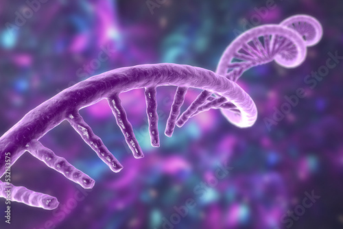
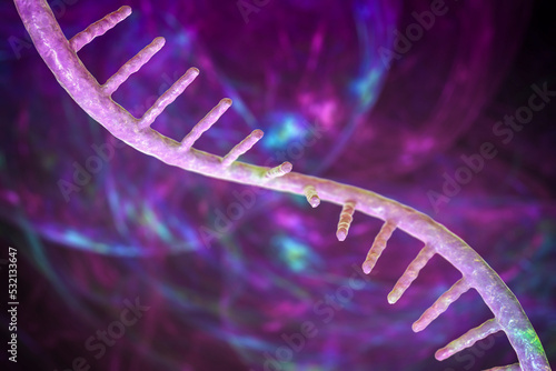
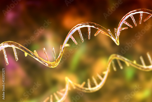
Molecule of mRNA, 3D illustration
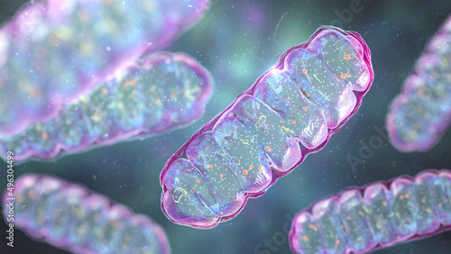
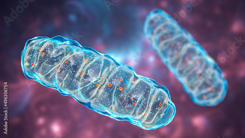
Mitochondria, a membrane-enclosed cellular organelles, which produce energy
Anterior cingulate cortex in human brain, 3D illustration.
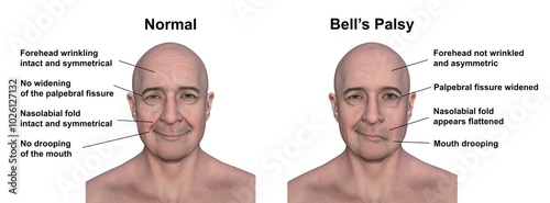
Facial palsy in a man, photorealistic 3D illustration
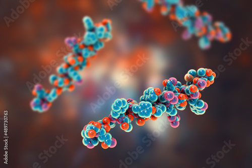
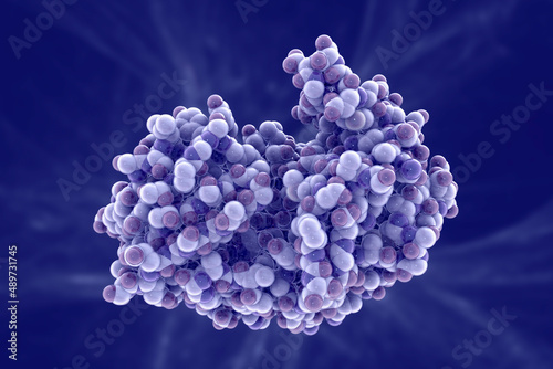
Molecule of the human hormone glucagon
Human skeleton in a ballet dancing pose, 3D illustration
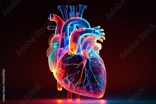
Anatomical model of human heart, ai illustration. Heart hologram
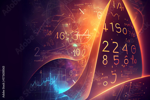
A fantasy math background featuring imaginary mathematical equations, formulas, and symbols, generative ai illustration
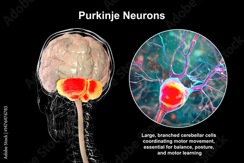
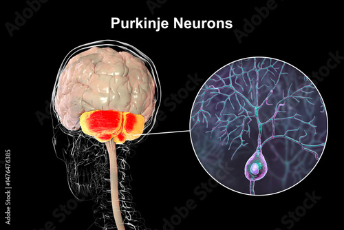
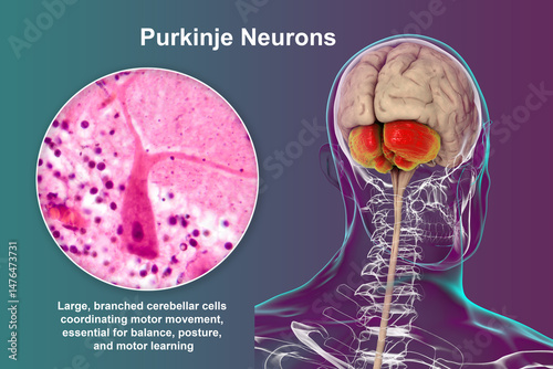
Purkinje neurons, 3D illustration showing large, branching neurons of the cerebellum responsible for motor coordination and balance control.
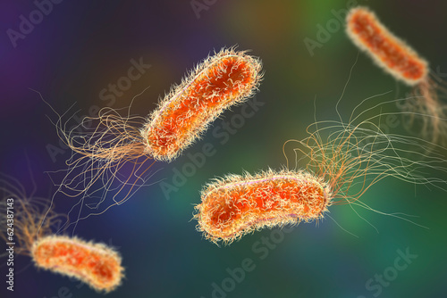
Pseudomonas aeruginosa bacteria, 3D illustration
Electroconvulsive therapy, ECT, a treatment involving the use of electrical currents to stimulate the brain, 3D illustration
Molecule of mRNA, 3D illustration
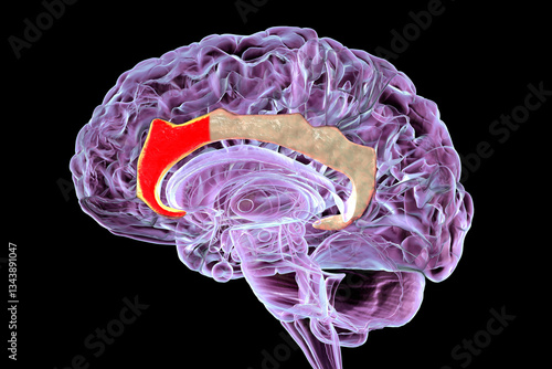
Corpus callosum and brain ventricles, 3D illustration
Molecule of mRNA, 3D illustration
A heartwarming illustration portraying people from diverse cultures coming together in a cooking, celebrating culinary traditions, unity, and cultural exchange, generative ai
Bifidobacterium bacteria, 3D illustration
Mitochondria, a membrane-enclosed cellular organelles, which produce energy
Molecule of pepsin stomach enzyme
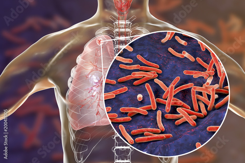
Secondary tuberculosis in lungs and close-up view of Mycobacterium tuberculosis bacteria, 3D illustration
Femur neck fracture types: subcapital, transcervical, and basicervical, 3D illustration
Cerebellum, 3D illustration and colored composite MRI
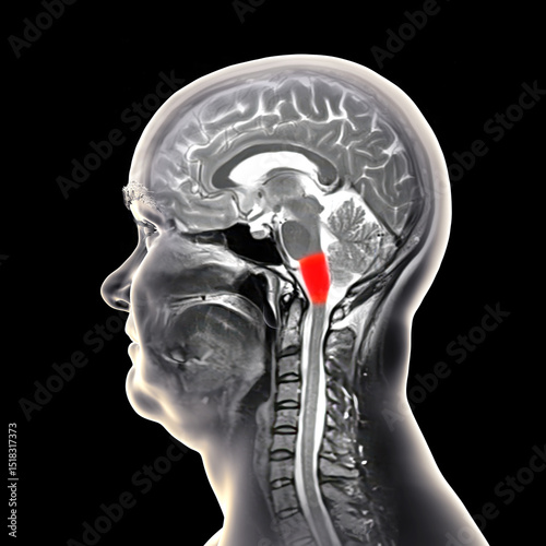
Medulla oblongata, 3D illustration and colored composite MRI
Medulla oblongata, 3D illustration and colored composite MRI
Cerebellum, colored composite MRI image and 3D illustration
Cerebellum, colored composite MRI image and 3D illustration
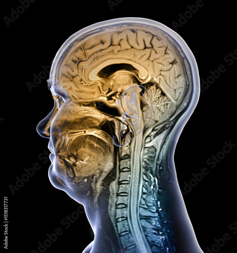
Brain anatomy, 3D illustration and composite MRI
Brain anatomy, 3D illustration and composite MRI
Brain anatomy, 3D illustration and composite MRI
Brain anatomy, 3D illustration and composite MRI
Medulla oblongata highlighted in human brain, colored composite MRI
Medulla oblongata highlighted in human brain, composite colored MRI and 3D illustration
Medulla oblongata highlighted in human brain, composite colored MRI and 3D illustration
Medulla oblongata highlighted in human brain, composite colored MRI and 3D illustration
Cerebellum, colored composite MRI image and 3D illustration
Generative AI illustration of a fantasy game interface featuring a vibrant green village landscape
Purkinje neurons, 3D illustration and light micrograph showing large, branching neurons of the cerebellar cortex, involved in motor coordination, and affected in ataxias.
Mirizzi syndrome, 3D illustration showing compression of the common hepatic duct by an impacted gallstone in the cystic duct, causing obstructive jaundice and inflammation.
Enteroviruses, 3D illustration showing spherical, non-enveloped RNA viruses responsible for diseases like meningitis, myocarditis, hand-foot-mouth disease.
Enteroviruses, 3D illustration showing spherical, non-enveloped RNA viruses responsible for diseases like meningitis, myocarditis, hand-foot-mouth disease.
Clostridium botulinum, 3D illustration. Bacterium producing botulinum toxin, leading to botulism with flaccid paralysis and respiratory failure.
Medulla oblongata highlighted in human brain, 3D illustration. Controls breathing, heartbeat, and other vital autonomic functions
Medulla oblongata highlighted in human brain, 3D illustration. Controls breathing, heartbeat, and other vital autonomic functions
Human brain with highlighted cerebellum, 3D illustration. Cerebellum is responsible for coordination, balance, and fine motor control. Damaged in ataxia and neurodegenerative disorders
Human brain with highlighted cerebellum and close-up of Purkinje neurons, 3D illustration and light micrograph.
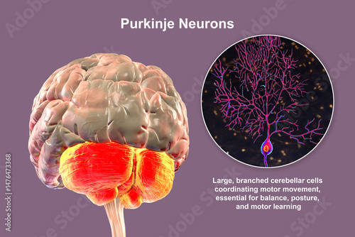
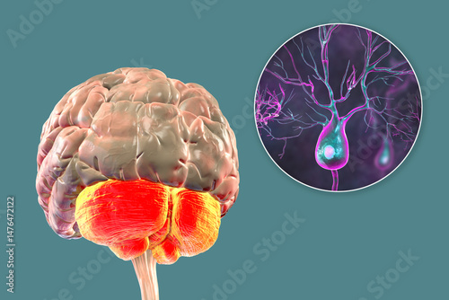
Human brain with highlighted cerebellum and close-up of Purkinje neurons, 3D illustration. Cerebellum controls balance and coordination, Purkinje cells are key neurons in motor function.
Human brain with highlighted cerebellum and close-up of Purkinje neurons, 3D illustration. Cerebellum controls balance and coordination, Purkinje cells are key neurons in motor function.
Human brain with highlighted cerebellum and close-up of Purkinje neurons, 3D illustration. Cerebellum controls balance and coordination, Purkinje cells are key neurons in motor function.
Human brain with highlighted cerebellum and close-up of Purkinje neurons, 3D illustration and light micrograph.
Human brain with highlighted cerebellum and close-up of Purkinje neurons, 3D illustration and light micrograph.
Human brain with highlighted cerebellum and close-up of Purkinje neurons, 3D illustration. Cerebellum controls balance and coordination, Purkinje cells are key neurons in motor function.
Human brain with highlighted cerebellum and close-up of Purkinje neurons, 3D illustration and light micrograph.
Human brain with highlighted cerebellum and close-up of Purkinje neurons, 3D illustration. Cerebellum controls balance and coordination, Purkinje cells are key neurons in motor function.
Human brain with highlighted cerebellum and close-up of Purkinje neurons, 3D illustration. Cerebellum controls balance and coordination, Purkinje cells are key neurons in motor function.
Human brain with highlighted cerebellum and close-up of Purkinje neurons, 3D illustration. Cerebellum controls balance and coordination, Purkinje cells are key neurons in motor function.
Human brain with highlighted cerebellum and close-up of Purkinje neurons, 3D illustration and light micrograph.
Human brain with highlighted cerebellum and close-up of Purkinje neurons, 3D illustration and light micrograph.
Human brain with highlighted cerebellum and close-up of Purkinje neurons, 3D illustration and light micrograph.
Human brain with highlighted cerebellum and close-up of Purkinje neurons, 3D illustration. Cerebellum controls balance and coordination, Purkinje cells are key neurons in motor function.
Human brain with highlighted cerebellum and close-up of Purkinje neurons, 3D illustration. Cerebellum controls balance and coordination, Purkinje cells are key neurons in motor function.
Human brain with highlighted cerebellum and close-up of Purkinje neurons, 3D illustration and light micrograph.
Human brain with highlighted cerebellum and close-up of Purkinje neurons, 3D illustration and light micrograph.
Human brain with highlighted cerebellum and close-up of Purkinje neurons, 3D illustration and light micrograph.
Human brain with highlighted cerebellum and close-up of Purkinje neurons, 3D illustration. Cerebellum controls balance and coordination, Purkinje cells are key neurons in motor function.
Human body anatomy with highlighted teeth, 3D illustration
Human body anatomy with highlighted teeth, 3D illustration
Human body anatomy with highlighted teeth, 3D illustration
Brain tumor, 3D illustration
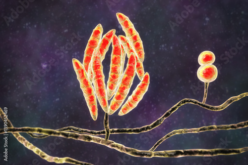
Fusarium fungi, 3D illustration
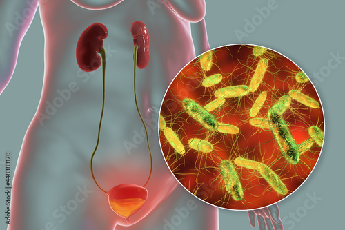
Cystitis, bacterial infection of urinary bladder, conceptual 3D illustration
Streptococcus pneumoniae bacteria, 3D scientific illustration
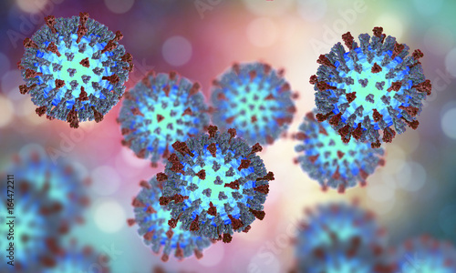
Measles viruses. 3D illustration showing structure of measles virus with surface glycoprotein spikes heamagglutinin-neuraminidase and fusion protein
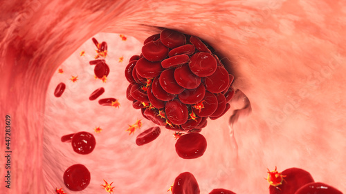
Blood clot in damaged blood vessel made of red blood cells, platelets and fibrin protein strands
Electroconvulsive therapy, ECT, a treatment involving the use of electrical currents to stimulate the brain, 3D illustration
Parasitic protozoans Toxoplasma gondii
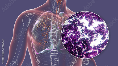
Lungs affected by silicosis, 3D illustration and micrograph
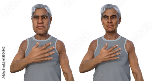
A man showing left-side stroke, 3D illustration
Mitochondria, a membrane-enclosed cellular organelles, which produce energy
Dengue viruses, 3D illustration
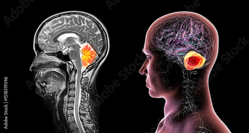
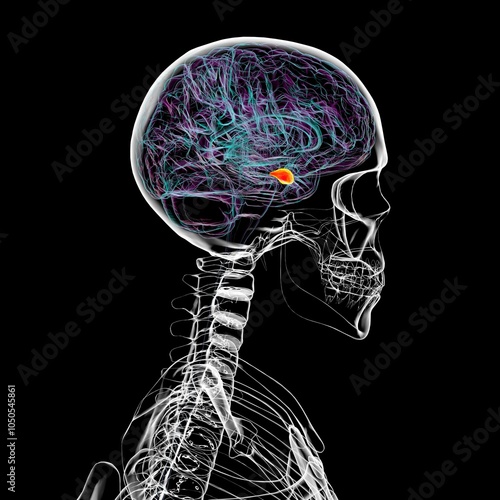
A pituitary gland tumor, 3D illustration
Human brain with the amygdala highlighted, 3D illustration
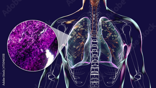
Lungs affected by silicosis, 3D illustration and micrograph
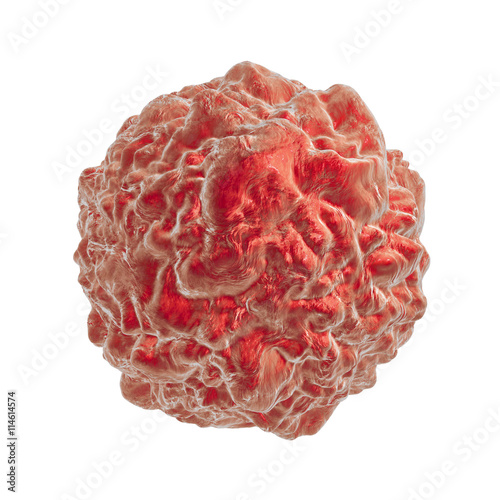
Tumor cell, cancer cell, carcinoma cell. 3D illustration
A doctor working in rehabilitation with a stroke patient during exercises, illustration
A man suffering from acromegaly, 3D illustration
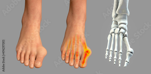
Toe deformation, also known as hallux valgus, or bunion