Login
Sign Up
Member Area
Login
Sign Up
English
Afrikaans formal
Afrikaans informal
Arabic
Azerbaijan
Brazilian
Bulgarian
Catalan
Chinese simplified
Chinese traditional
Croatian
Czech
Danish
Dutch
English
Estonian
Finnish
French
Georgian
German
Greek
Hebrew
Hungarian
Icelandic
Indonesian
Italian
Japanese
Latvian
Lithuanian
Malaysian
Norwegian
Persian
Polish
Portuguese
Romanian
Russian
Serbian
Slovakian
Slovenian
Spanish
Swedish
Thai
Turkish
Ukrainian
Vietnamese
Toggle navigation
VideotonStock
Your Shopping Cart is empty.
Files
Stock
Photo
Video
Special offer
Collections
Packages
Lightboxes
Licenses
Royalty free
Rights managed
Creative
Editorial
Items
Most downloaded
Featured
Most popular
New
Free
Categories
Browse categories
Site info
Site info
About
Support
Privacy Policy
Privacy Policy
FAQ
Contact Us
Contact Us
Terms and Conditions
Buyer Agreement
Seller Agreement
Customers
Users
Credits
Photographers
Boby Richards
0
Boby Richards
0
Videoton1
286
Search
Keywords:
Creator ID:
Type:
Photo
Illustration
Vector
Video
Template
3d
Categories:
Animals
Buildings and Architecture
Business
Drinks
The Environment
States of Mind
Food
Graphic Resources
Hobbies and Leisure
Industry
Landscapes
Lifestyle
People
Plants and Flowers
Culture and Religion
Science
Social Issues
Sports
Technology
Transport
Travel
News
Entertainment
Sport News
Languages:
Brazilan Portuguese - Brazil
English - Canada
French - Canada
Spanish - Mexico
English - United States
English - Belgium
French - Belgium
Dutch - Belgium
Czech - Czech Republic
English - Cyprus
Danish - Danmark
German - Germany
English - Estonia
Spanish - Spain
French - France
French - Marocco
English - Greece
English - Ireland
Italian - Italia
English - Latvia
English - Lithuania
German - Luxembourg
English - Luxembourg
French - Luxembourg
English - Hungary
English - Malta
Dutch - Nederlands
Bokmal Norwegian - Norway
German - Austria
Polish - Poland
Portuguese - Portugal
English - Romania
German - Switzerland
English - Slovenia
Slovakian - Slovakia
French - Switzerland
English - Finland
Swedish- Sweden
Italian - Switzerland
English - United Kingdom
English - Bulgaria
English - Australia
Japanese - Japan
Korean - South Korea
English - New Zealand
Russian - Russia
English - Ukrain
English - Thailand
Orientation:
All
Horizontal
Vertical
square
Color:
Model property release:
All
Yes
No
Duration Video:
All
Up to 10 seconds
Up to 20 seconds
Longer than 30 seconds
Results
(0)
Site
Getty/iStock
Bigstockphoto
Pixabay
Adobe Stock
Relevance
New
Most popular
Most downloaded
Undiscovered
12
24
36
48
96
Auto paging
Menu
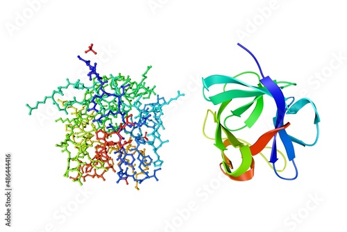
Insulin, monoclinic crystal form. Ribbons diagram with differently colored protein chains based on protein data bank entry 1znj. Scientific background. 3d illustration
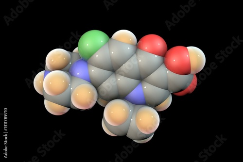
Molecular structure of ciprofloxacin antibiotic, 3D illustration
Human receptor-interacting protein kinase 1 domain in complex with a novel inhibitor. Rendering with differently colored protein chains based on protein data bank entry 7fd0. 3d illustration
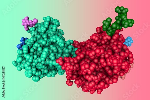
Human estrogen-related receptor gamma ligand binding domain complex with bisphenol Z. Crystal structure and molecular models. Rendering based on protein data bank. Rainbow coloring from N to C. 3d
Structure of Herceptin Fab without antigen. Ribbons diagram with differently colored protein chains based on protein data bank entry 6mh2. 3d illustration
Recombinant human calcyphosine, a novel EF-hand-containing protein. Molecular model. Rendering based on protein data bank entry 3e3r. Scientific background. 3d illustration
X-ray structure of human thioredoxin reductase 1. Ribbons diagram with differently colored protein chains based on protein data bank. Scientific background. 3d illustration
Human beta-defensin-2. Molecular model on blue background. Rendering based on protein data bank entry 1fd3. Scientific background. 3d illustration
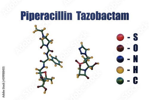
Piperacillin and tazobactam, a combination medication containing rhe antibiotic piperacillin and the beta-lactamase inhibitor tazobactam. Molecular models isolated on white background. 3d illustration
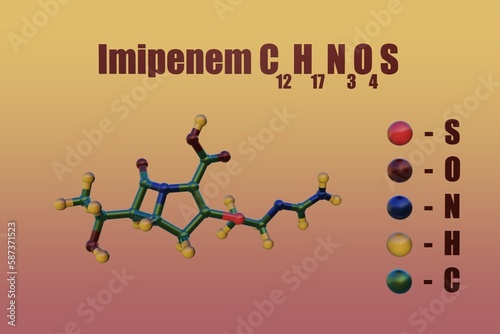
Structural chemical formula and molecular model of imipenem, an ultravenous beta-lactam antibiotic belong to the subgroup of carbapenems. 3d illustration
Human erythropoietin, protein that stimulates the production of red blood cells. Rendering based on protein data bank entry 1buy. Rainbow coloring from N to C. 3d illustration
Crystal structure of human intelectin-1 or omentin. Ribbons diagram in very peri coloring. Rendering based on protein data bank entry 4wmq. Scientific background. 3d illustration
Space-filling molecular model of ipidacrine or amiridine. Atoms are represented as spheres with conventional color coding: carbon (grey), nitrogen (blue), hydrogen (white). 3d illustration
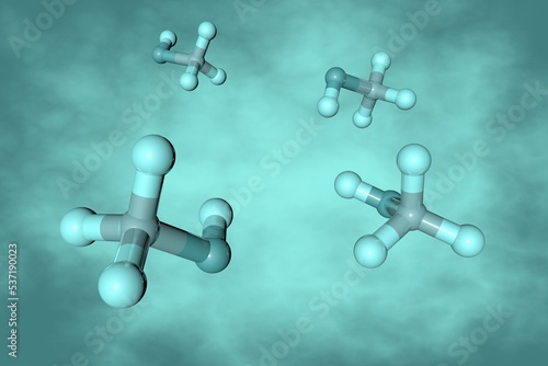
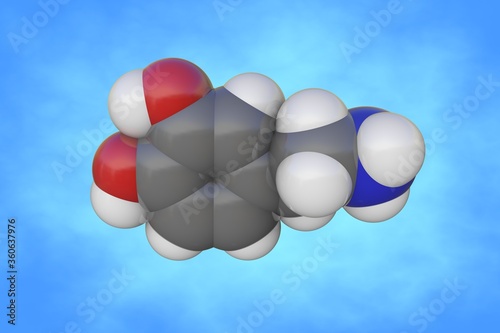
Space-filling molecular model of methylamine. Atoms are represented as spheres with conventional color coding: carbon (grey), nitrogen (blue), hydrogen (white). 3d illustration
Molecular model of human methionine aminopeptidase-2, an enzyme that in humans is encoded by the METAP2 gene. Scientific background. 3d illustration
Crystal structure and space-filling molecular model of human interleukin 1 beta. Rendering based on protein data bank. Rainbow coloring from N to C. Scientific background. 3d illustration
Crystal structure and molecular model of deoxyhemoglobin in complex with aryloxyalkanoic acid. Rendering based on protein data bank. Rainbow coloring from N to C. 3d illustration
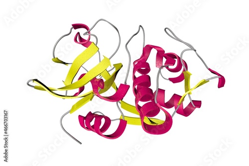
Solution structure of human interleukin-33. Ribbons diagram in secondary structure coloring based on protein data bank entry 2kll. Scientific background. 3d illustration
Molecular model of human interleukin-33 isolated on white background. Rendering based on protein data bank entry 2kll. Rainbow coloring from N to C. 3d illustration
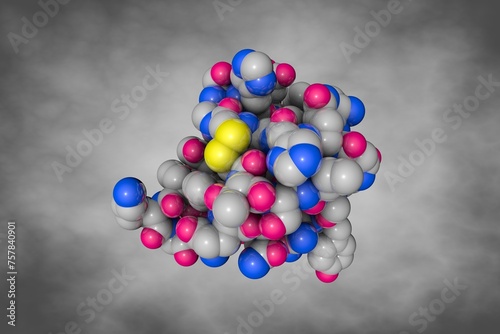
Space-filling molecular model of human interleukin-33. Atoms are shown as spheres with color coding: carbon (grey), oxygen (red), hydrogen (white), nitrogen (blue), sulfur (yellow). 3d illustration
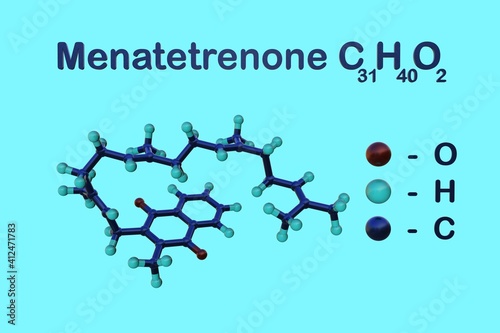
Structural chemical formula and molecular model of menatetrenone, one of the nine forms of vitamin K2. It is produces by converting vitamin K1 in the body, testicles, and pancreas. 3d illustration
Crystal structure of human dipeptidyl peptidase-4, a serine protease, a member of the prolyl oligopeptidase family that has been implicated in several diseases. 3d illustration
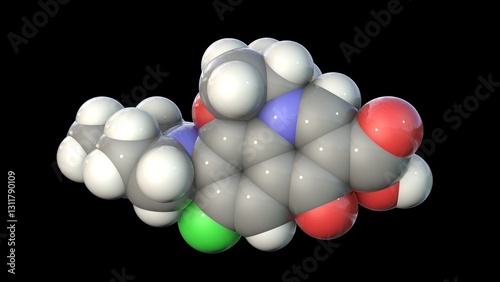
Molecular structure of levofloxacin, a fluoroquinolone antibiotic, 3D illustration
Molecular structure of the N-terminal domain of carcinoembryonic antigen (CEA). Rendering based on protein data bank entry 2qsq. Scientific background. 3d illustration
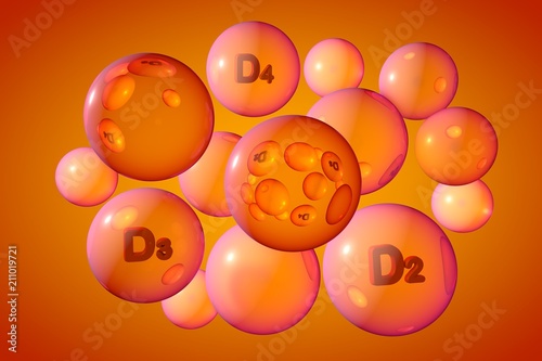
Transparent orange vitamin D2, D3, D4 pills. Ergocalciferol, cholecalciferol and dihydroergocalciferol capsules. Vitamin and mineral complex. 3d illustration
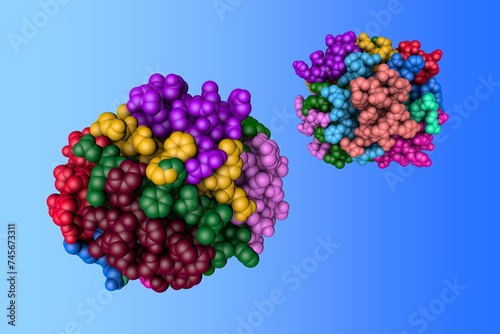
Molecular model of alpha-galactosidase, enzyme that associated with Fabry disease. Rendering with multi-colored protein chains based on protein data bank. 3d illustration
Space-filling molecular model of human brain-type creatine kinase, a transferase protein belonging to the phosphagen kinase superfamily. 3d illustration
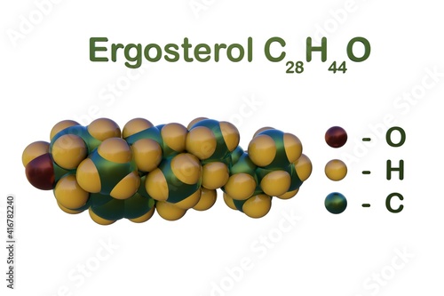
Structural chemical formula and space-filling molecular model of ergosterol or provitamin D2, a sterol found in cell membranes of fungi and protozoa. 3d illustration
Human hemoglobin nitric oxide adduct. Ribbons diagram in secondary structure colors based on protein data bank entry 4n8t. 3d illustration
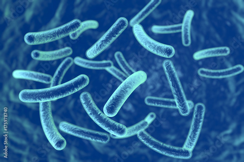
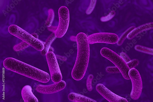
Bacteria on blue background, 3D illustration
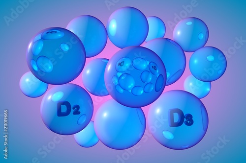
Transparent blue vitamin D2, D3 pills. Ergocalciferol and cholecalciferol capsules. Vitamin and mineral complex. 3d illustration
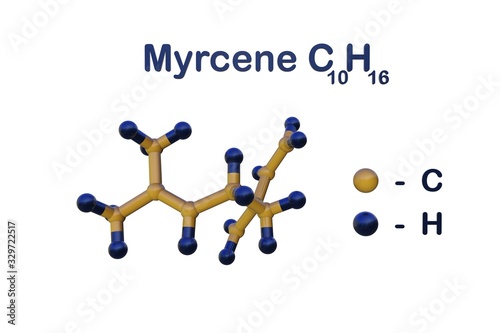
Structural chemical formula and molecular model of myrcene, the most abundant terpene in modern commercial cannabis. Scientific background. 3d illustration
Crystal structure of human sonic hedgehog N-terminal domain. Ribbons diagram in secondary structure coloring based on protein data bank entry 3m1n. 3d illustration
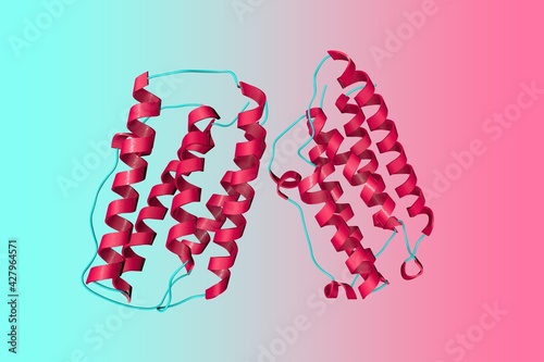
Structure of cell wall targeting domain of peptidylglycan hydrolase ALE-1. Ribbons diagram with differently colored protein chains based on protein data bank entry 1r77. 3d illustration
Human S-adenosyl methionine synthetase 1 in complex with SAM and PPNP. Ribbons diagram on purple background. Rendering based on protein data bank entry 8swa. 3d illustration
Human progesterone receptor ligand binding domain in complex with the ligand metribolone. Molecular model. Rendering based on protein data bank entry 1e3k. Scientific background. 3d illustration
Structural chemical formula and space-filling molecular model of mirtazapine, an antidepressant medicine used to treat depression, compulsive and anxiety disorders. 3d illustration
Human serum albumin complexed with hexadecanoic acid (palmitic acid). Ribbons diagram based on protein data bank entry 1e7h. Rainbow coloring from N to C. 3d illustration
Portrait of mystical dragon, watercolor style, generative ai illustration
Human recombinant Gla-domainless prothrombin mutant. Space-filling molecular model. Rendering with differently colored protein chains based on protein data bank entry 4hzh. 3d illustration
Human recombinant Gla-domainless prothrombin mutant. Ribbons diagram with differently colored protein chains based on protein data bank entry 4hzh. 3d illustration
Human fatty acid synthase. Space-filling molecular model. Rainbow coloring from N to C. 3d illustration
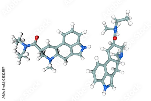
Molecular structure of levofloxacin, a fluoroquinolone antibiotic, 3D illustration
Molecular structure of levofloxacin, a fluoroquinolone antibiotic, 3D illustration
Molecular structure of levofloxacin, a fluoroquinolone antibiotic, 3D illustration
Molecular structure of ciprofloxacin antibiotic, 3D illustration
Molecular structure of ciprofloxacin antibiotic, 3D illustration
Molecular structure of ciprofloxacin antibiotic, 3D illustration
Molecular structure of ciprofloxacin antibiotic, 3D illustration
Molecular model of human interleukin-6 or IL-6. Scientific background. 3d illustration
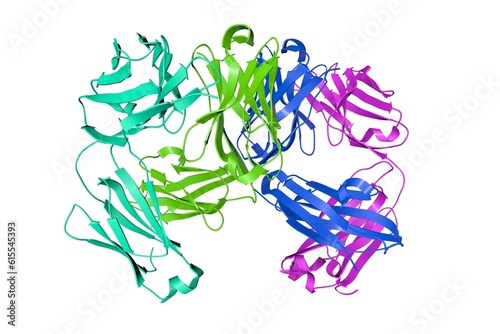
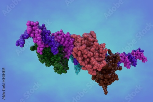
Crystal structure and space-filling molecular model of the intact human immunoglobulin isolated on white background. Rendering with differently colored protein chains. 3d illustration
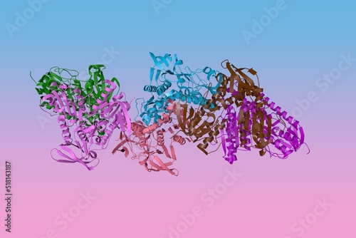
Influenza A virus H7N9 polymerase apo-protein dimer complex. Ribbons diagram with differently colored protein chains based on protein data bank entry 7zpm. Scientific background. 3d illustration
Methanol or methyl alcohol. Molecular model on turquoise background. Scientific background. 3d illustration
Acetaldehyde or ethanal, an organic chemical compound produced by plants and one of the most important aldehydes. Molecular model on blue background. Scientific background. 3d illustration
Structural chemical formula and space-filling molecular model of hesperidin, a bioactive flavonoid that possess antioxidant, anti-carcinogenic and other therapeutic properties. 3d illustration
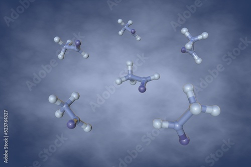
Space-filling molecular model of histamine. It is an organic nitrogenous compound involved in local immune responses and acting as a neurotransmitter. 3d illustration
Crystal structure and molecular model of fibroblast growth factor 4 isolated on white background. Rendering based on protein data bank entry 1ijt. Rainbow coloring from N to C. 3d illustration
Molecular model of sugar binding protein - human galectin-2 (dimer) on colorful background. Rendering based on protein data bank. Scientific background. 3d illustration
Bacteria on colourful background, 3D illustration
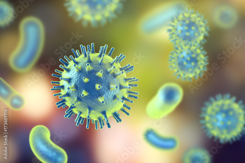
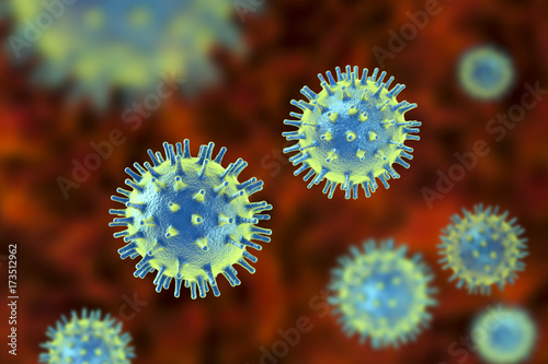
Human pathogenic viruses and bacteria, 3d illustration
Crystal structure and molecular model of human intelectin-1, also known as omentin. Rendering with differently colored protein chains based on protein data bank. 3d illustration
Crystal structure of human cathepsin K, an enzyme that in humans is encoded by the CTSK gene. Ribbons diagram in secondary structure coloring based on protein data bank. 3d illustration
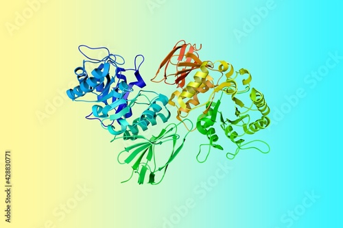
Crystal structure of human pancreatic alpha-amylase in complex with the mechanism based inactivator glucosyl cyclophellitol. 3d illustration
Molecular model of human pancreatic alpha-amylase. Atoms are shown as color-coded spheres: carbon (gray), hydrogen (white), oxygen (red), nitrogen (blue), sulfur (yellow). 3d illustration
Regulator of chromosome condensation of human (RCC1). Rendering with differently colored protein chains based on protein data bank. Scientific background. 3d illustration
Space-filling molecular model of human intelectin-1. Scientific background. 3d illustration
Molecular model of interferon regulating factor 3 bound to the interferon-beta enhancer. Rendering with differently colored protein chains based on protein data bank. 3d illustration
Crystal structure of alpha-galactosidase enzyme that causes Fabry disease. Ribbons diagram in rainbow colors. Scientific background. 3d illustration
Crystal structure of human interferon beta, a cytokine in the interferon family used to treat multiple sclerosis. 3d illustration
Structural chemical formula and space-filling molecular model of rutin, a bioflavonoid found in certain vegetables and fruits that has powerful antioxidant properties. 3d illustration
Structural chemical formula and molecular model of mandelic acid, an alpha hydroxy acid derived from almond. It is an anti-ageing skin ingredient. 3d illustration
Molecular structure of dobutamine, a medication used in the treatment of cardiogenic shock and severe heart failure. Scientific background. 3d illustration
Space-filling molecular model of norepinephrine. Atoms are represented as spheres with color coding: carbon (grey), oxygen (red), nitrogen (blue), hydrogen (white). 3d illustration
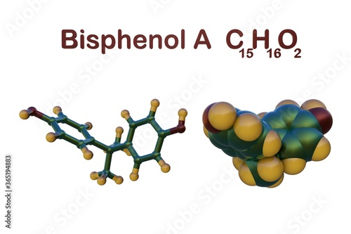
Structural chemical formula and molecular model of bisphenol A, a chemical produced for use in the production of polycarbonate plastics and epoxy resins. 3d illustration
Molecular structure of glycerol. Atoms are represented as spheres with color coding: carbon (grey), oxygen (red), hydrogen (white). 3d illustration
Hepatitis C viruses in blood, 3D illustration
Space-filling molecular model of the insulin receptor ectodomain in complex with one insulin molecule isolated on white background. Medical background. Scientific background. 3d illustration
Hepatitis C viruses on red background, 3D illustration
Rhinovirus isolated on black background, 3D illustration
Earth from space
Molecular structure of lysergide (LSD). Atoms are represented as spheres with conventional color coding: hydrogen (white), carbon (grey), oxygen (red), nitrogen (blue). 3d illustration
Molecular structure of curcumin, molecule produced by turmeric roots. Atoms are represented as spheres with color coding: oxygen (red), hydrogen (green), carbon (yellow). 3d illustration
Colorful virus on purple background. 3d illustration
Hepatitis A virus isolated on black background, 3D illustration
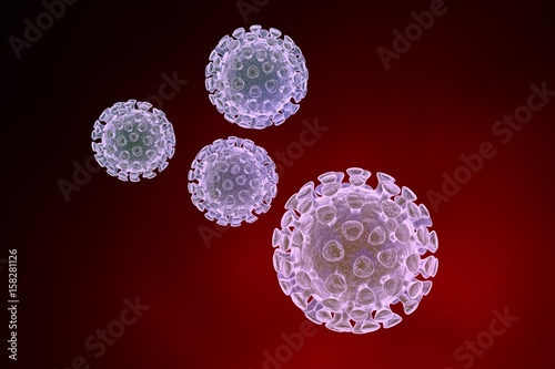
Human pathogenic viruses, 3D illustration
Structure of human sonic hedgehog N-terminal domain. Space-filling molecular model with differently colored protein chains based on protein data bank entry 3m1n. 3d illustration
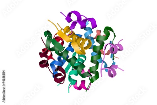
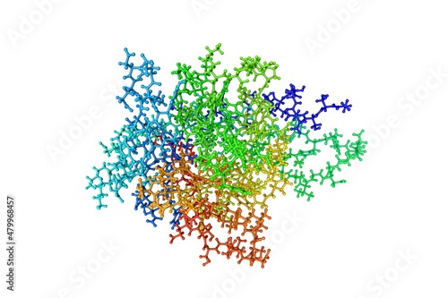
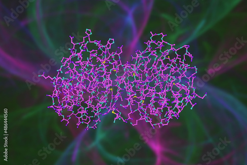
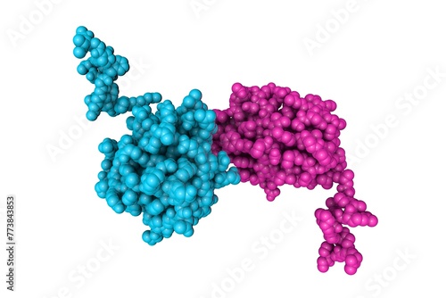
Human insulin. Crystal structure and molecular model. Rendering with differently colored protein chains based on protein data bank entry 3i3z. Scientific background. 3d illustration
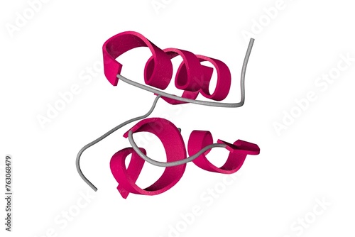
Human sonic hedgehog N-terminal domain. Molecular model based on protein data bank entry 3m1n. Scientific background. 3d illustration
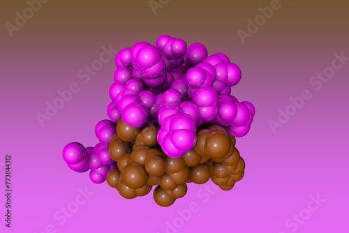
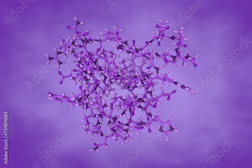
Human insulin. Space-filling molecular model on blue background. Rendering based on protein data bank entry 3i3z. Scientific background. 3d illustration
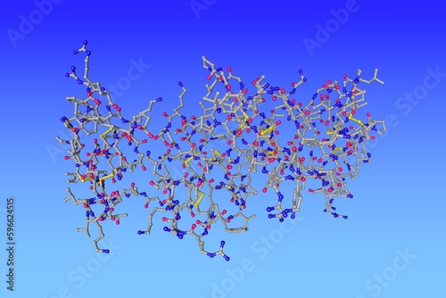
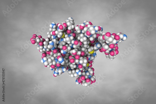
Human insulin. Space-filling molecular model. 3d illustration
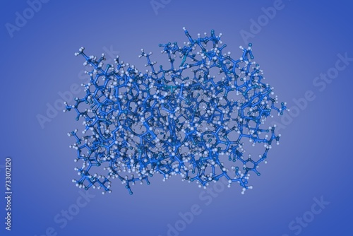
Human insulin. Space-filling molecular model on gray background. Rendering based on protein data bank entry 3i3z. Scientific background. 3d illustration
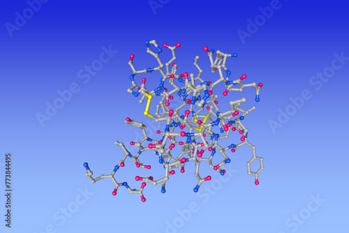
Human insulin. Ribbons diagram in secondary structure coloring based on protein data bank entry 3i3z. Scientific background. 3d illustration
Crystal structure of human sonic hedgehog N-terminal domain. Ribbons diagram with differently colored protein chains based on protein data bank entry 3m1n. 3d illustration
Structure of human sonic hedgehog N-terminal domain. Molecular model with differently colored protein chains based on protein data bank entry 3m1n. 3d illustration
Insulin, monoclinic crystal form. Space-filling molecular model. Rendering with differently colored protein chains based on protein data bank entry 1znj. Scientific background. 3d illustration
Structure of human sonic hedgehog on orange background. Ribbons diagram based on protein data bank entry 6pjv. Scientific background. 3d illustration
NMR Structure of single-chain insulin on very peri background. Molecular model based on protein data bank entry 2lwz. Scientific background. 3d illustration
Structure of the cell wall targeting domain of peptidylglycan hydrolase ALE-1. Space-filling molecular model. Rendering based on protein data bank entry 1r77. Scientific background. 3d illustration
Structure of human sonic hedgehog in complex with zinc and magnesium on blue background. Molecular model based on protein data bank entry 6pjv. 3d illustration
Single-chain insulin. Space-filling molecular model. Atoms are shown as spheres with color coding: carbon (grey), oxygen (red), hydrogen (white), nitrogen (blue), sulfur (yellow). 3d illustration