Login
Sign Up
Member Area
Login
Sign Up
English
Afrikaans formal
Afrikaans informal
Arabic
Azerbaijan
Brazilian
Bulgarian
Catalan
Chinese simplified
Chinese traditional
Croatian
Czech
Danish
Dutch
English
Estonian
Finnish
French
Georgian
German
Greek
Hebrew
Hungarian
Icelandic
Indonesian
Italian
Japanese
Latvian
Lithuanian
Malaysian
Norwegian
Persian
Polish
Portuguese
Romanian
Russian
Serbian
Slovakian
Slovenian
Spanish
Swedish
Thai
Turkish
Ukrainian
Vietnamese
Toggle navigation
VideotonStock
Your Shopping Cart is empty.
Files
Stock
Photo
Video
Special offer
Collections
Packages
Lightboxes
Licenses
Royalty free
Rights managed
Creative
Editorial
Items
Most downloaded
Featured
Most popular
New
Free
Categories
Browse categories
Site info
Site info
About
Support
Privacy Policy
Privacy Policy
FAQ
Contact Us
Contact Us
Terms and Conditions
Buyer Agreement
Seller Agreement
Customers
Users
Credits
Photographers
Boby Richards
0
Boby Richards
0
Videoton1
286
Search
Keywords:
Creator ID:
Type:
Photo
Illustration
Vector
Video
Template
3d
Categories:
Animals
Buildings and Architecture
Business
Drinks
The Environment
States of Mind
Food
Graphic Resources
Hobbies and Leisure
Industry
Landscapes
Lifestyle
People
Plants and Flowers
Culture and Religion
Science
Social Issues
Sports
Technology
Transport
Travel
News
Entertainment
Sport News
Languages:
Brazilan Portuguese - Brazil
English - Canada
French - Canada
Spanish - Mexico
English - United States
English - Belgium
French - Belgium
Dutch - Belgium
Czech - Czech Republic
English - Cyprus
Danish - Danmark
German - Germany
English - Estonia
Spanish - Spain
French - France
French - Marocco
English - Greece
English - Ireland
Italian - Italia
English - Latvia
English - Lithuania
German - Luxembourg
English - Luxembourg
French - Luxembourg
English - Hungary
English - Malta
Dutch - Nederlands
Bokmal Norwegian - Norway
German - Austria
Polish - Poland
Portuguese - Portugal
English - Romania
German - Switzerland
English - Slovenia
Slovakian - Slovakia
French - Switzerland
English - Finland
Swedish- Sweden
Italian - Switzerland
English - United Kingdom
English - Bulgaria
English - Australia
Japanese - Japan
Korean - South Korea
English - New Zealand
Russian - Russia
English - Ukrain
English - Thailand
Orientation:
All
Horizontal
Vertical
square
Color:
Model property release:
All
Yes
No
Duration Video:
All
Up to 10 seconds
Up to 20 seconds
Longer than 30 seconds
Results
(0)
Site
Getty/iStock
Bigstockphoto
Pixabay
Adobe Stock
Relevance
New
Most popular
Most downloaded
Undiscovered
12
24
36
48
96
Auto paging
Menu
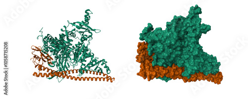
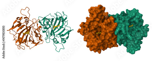
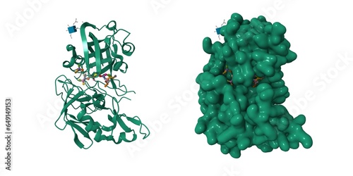
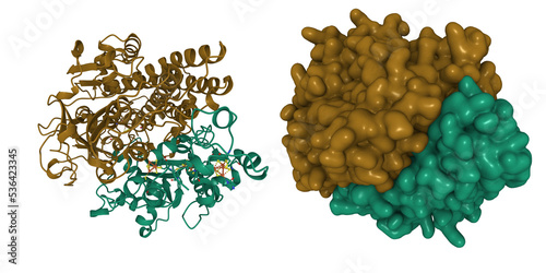
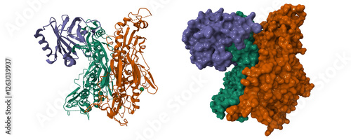
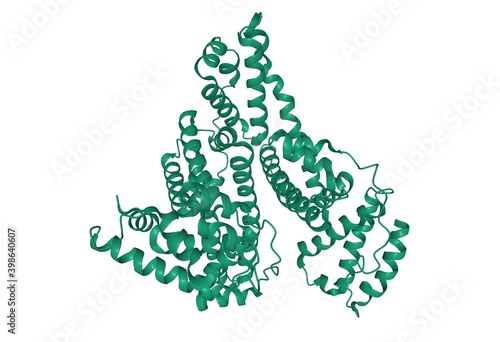
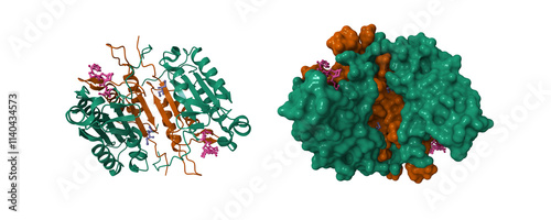
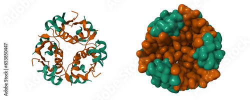
Crystal structure of PI3Kalpha. Catalytic subunit is shown in green, regulatory subunit im brown. 3D cartoon and molecular surface models, PDB7pg5, entity id color scheme
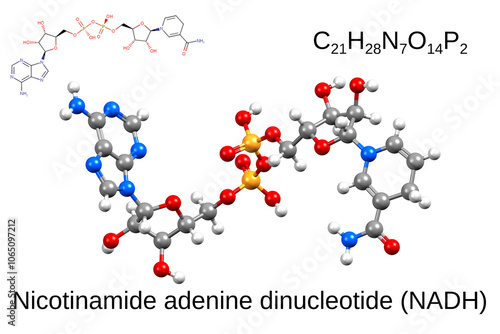
Structure of nicotinamide adenine dinucleotide. Chemical formula, skeletal formula, and 3D ball-and-stick model
Structure of the human collapsin response mediator protein-1, a lung cancer suppressor. 3D cartoon and molecular surface models, chain id color scheme, pdb 4b3z
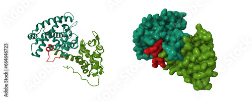
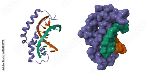
Crystal structure of the retinoblastoma tumour suppressor protein (green) bound to E2F peptide. 3D cartoon and Gaussian surface models, PDB 1o9k, chain id color scheme
Structure of botulinum neurotoxin serotype A, 3D cartoon model isolated, secondary structure color scheme, PDB 3bta
Activated spinach rubisco in complex with the product 3-phosphoglycerate (pink). 3D cartoon and molecular surface model, PDB 1aa1
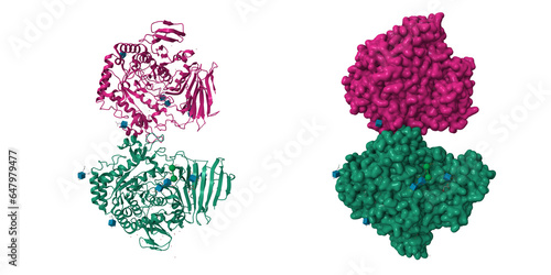
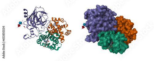
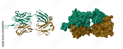
Crystal structure of the dimeric extracellular domain of human carbonic anhydrase XII with inhibitor. 3D cartoon and molecular surface models, chain id color scheme, pdb 1jd0
Structure of the progesterone receptor-DNA complex, 3D cartoon model, PDB 2c7a
Crystal structure of the MEILB2-BRCA2 complex. 3D cartoon and molecular surface models, chain id color scheme, pdb 7ldg
Crystal structure of free ERI2 nuclease. 3D cartoon and molecular surface models, chain id color scheme, pdb 7n8v
Cryo-EM structure of c9orf72 (violet)-SMCR8 (brown)-WDR41 (green). 3D cartoon and molecular surface models, pdb 6lt0
High resolution crystal structure of substrate-free human neprilysin. 3D cartoon model, PDB 6gid
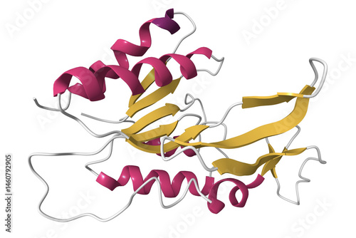
Crystal structure of human saposin A. 3D cartoon model, PDB 1n69, secondary structure color scheme
Crystal structure of human cytochrome P450 3A4. 3D cartoon model, PDB 1w0e, secondary structure color scheme, white background.
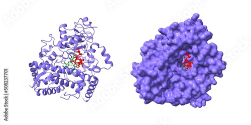
Crystal structure of renin with inhibitor aliskiren. 3D cartoon and Gaussian surface models, chain id color scheme, PDB 2v0z
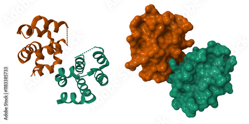
Crystal structure of oxidized alpha hemoglobin (green) bound to alpha-haemoglobin-stabilizing protein. 3D cartoon and Gaussian surface models, chain id color scheme, PDB 1z8u
Crystal structure of human P450 3A4 in complex with erythromycin (red). The protoporphyrin is shown in green. 3D cartoon and Gaussian surface models, PDB 2j0D, white background.
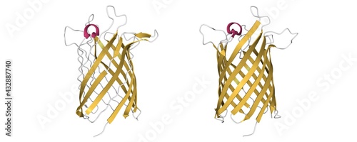
Structure of the monomeric outer membrane porin OmpG in the open (left) and closed (right) conformation, 3D cartoon model with differently colored secondary structure elements, white background
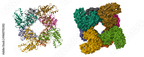
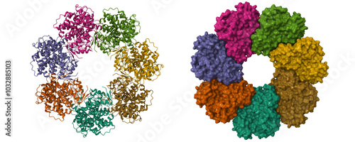
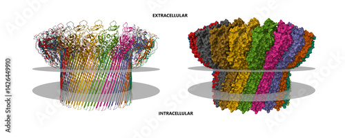
Cryo-EM structure of the active NLRP3 inflammasome disk. 3D cartoon and Gaussian surface models, PDB 8ej4, entity id color scheme
Crystal structure of unliganded myogenesis-regulating glycosidase. 3D cartoon and Gaussian surface models, PDB 7qqf, white background
Crystal structure of cathepsin A, apo-structure. 3D cartoon and Gaussian surface models, chain id color scheme, PDB 4ci9, white background
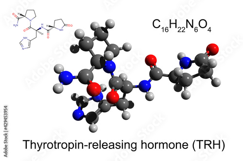
Skeletal and chemical formulas, and 3D ball-and-stick model of the human thyrotropin-releasing hormone, white background
Chemical formula, skeletal formula and 3D ball-and-stick model of propofol
Human erythropoietin, 3D cartoon model isolated
Crystal structure of the oxidized form of Ni-Fe hydrogenase of Desulfovibrio gigas. 3D cartoon and Gaussian surface models, entity id color scheme, PDB 2frv
Chemical formula, structural formula and 3D ball-and-stick model of aliskiren, a direct renin inhibitor
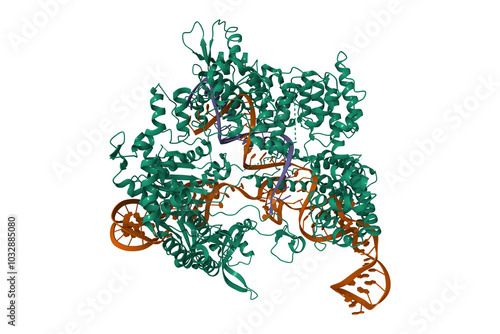
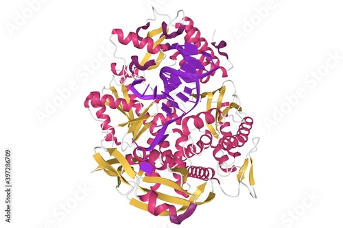
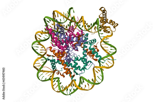
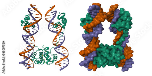
Crystal structure of Streptococcus pyogenes Cas9 in complex with guide RNA (brown) and target DNA (violet). 3D cartoon model, PDB 4oo8
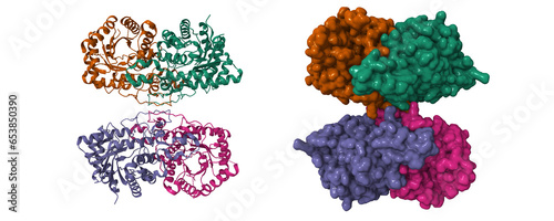
Crystal structure of human complement component C8. 3D cartoon and molecular surface models, chain id color scheme, pdb 3ojy
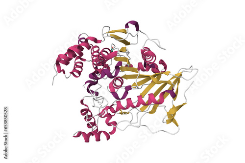
Solution structure of a human cancer-related nucleoside triphosphatase. 3D cartoon model, secondary structure color scheme, pdb 2i3b
Structure of human mitochondrial peptide deformylase, a novel cancer target. 3D cartoon and molecular surface models, chain id color scheme, pdb 3g5b
Cryo-EM structure of human uMtCK1. 3D cartoon and molecular surface models, chain id color scheme, pdb 9b05
Solution structure of human guanylate kinase. 3D cartoon model, secondary structure color scheme, pdb 6nui
Crystal structure of human glutaminase kidney isoform in complex with inhibitor. 3D cartoon and molecular surface models, chain id color scheme, pdb 6ukb
Crystal structure of Mcl1 with inhibitor. 3D cartoon model, secondary structure color scheme, pdb 6oqd
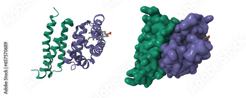
G2-mitotic-specific cyclin B1 (pink), cyclin-dependent kinase 1 (blue) and cyclin-dependent kinase regulatory subunit 2 (green) with Flavopiridol. 3D cartoon and Gaussian surface models, PDB 6gu2
The crystal structure of the isolated streptokinase middle (SKbeta) domain. 3D cartoon and molecular surface models, chain id color scheme, PDB 1c4p
Cryo-EM structure of human hormone thyroglobulin. 3D cartoon and molecular surface models, PDB 7b75
Structure of human albumin, 3D cartoon model isolated, white background
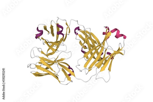
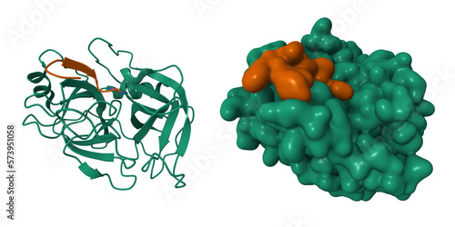
Structure of the Fab region of cetuximab, an anticancer drug. 3D cartoon model, secondary structure color scheme, PDB 5sx4, white background
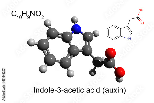
Chemical formula, skeletal formula, and 3D ball-and-stick model of indole-3-acetic acid, the most common plant auxin, white background
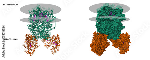
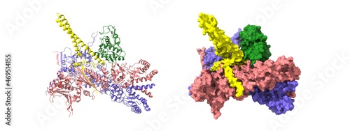
Cryo-EM structure of the human TRPC5 ion channel (green) in complex with G alpha i3 subunits (brown), class1. 3D cartoon and molecular surface models, PDB 7x6i
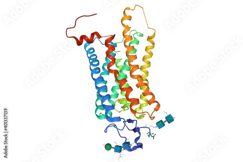
Crystal structure of photoactivated rhodopsin, 3D cartoon model isolated, white background
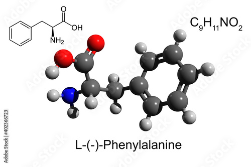
Chemical formula, skeletal formula and 3D ball-and-stick model of L-phenylalanine, an essential amino acid, white background
Heat-shock protein 90 dimer (pink-blue)-HSP90 co-chaperone Cdc37 (yellow)-cyclin-dependent kinase 4 (green) complex. 3D cartoon and Gaussian surface models, PDB 5fwk, white background
Structure of human haptoglobin-haemoglobin complex. 3D cartoon and Gaussian surface models, chain id color scheme, PDB 4x0l
Pyrococcus abyssi B family DNA polymerase bound to a dsDNA, 3D cartoon model isolated, white background
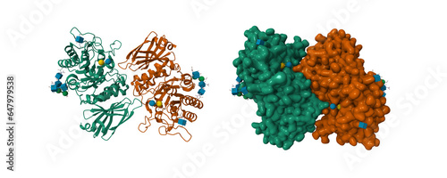
Crystal structure of human galectin-1 in complex with type 1 N-acetyllactosamine. 3D cartoon and Gaussian surface model, chain id color scheme, PDB 4xbl, white background
Crystal structure of human 2909 Fab, a quaternary structure-specific antibody against HIV-1. 3D cartoon and molecular surface models, chain id color scheme, pdb 3piq
Structure of human interleukin-23 heterodimer, 3D cartoon model isolated, white background
Crystal structure of Bacillus thuringiensis Cry5B nematocidal toxin, 3D cartoon and Gaussian surface models, chain instance color scheme, based on PDB 4d8m, white background
Caspase-1 complex with interleukin-18 (violet). 3D cartoon and molecular surface models, PDB 8sv1, entity id color schem
Dimeric structure of CARMA1 CARD. 3D cartoon and molecular surface models, PDB 4jup, chain id color scheme
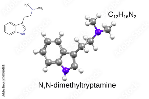
Chemical formula, structural formula and 3D ball-and-stick model of N,N-Dimethyltryptamine
Caspase 8 bound to a covalent inhibitor (pink). 3D cartoon and molecular surface models, PDB 3kjn, chain id color scheme
Crystal structure of human ornithine transcarbamylase complexed with carbamoyl phosphate. 3D cartoon and molecular surface models, PDB 1fvo, chain instance color scheme
Chemical formula, structural formula and 3D ball-and-stick model of L-arginine, an essential amino acid, white background
DNA polymerase beta (POLB) complexed with seven base pairs of DNA and deoxythymidine triphosphate (dTTP), 3D cartoon model in two perpendicular projections, white background
Crystal structure of native recombinant human bile salt activated lipase. 3D cartoon model, secondary structure color scheme, PDB 6h0t
Cryo-EM structure of retinoblastoma-binding protein 5 (green) bound to the nucleosome. 3D cartoon and Gaussian surface models, PDB 6pwx
Structure of human interleukin-33, 3D cartoon model isolated with differently colored elements of the secondary structure, white background
Crystal structure of human thymidine kinase 1 active center with bound thymidine triphosphate. 3D cartoon model, PDB 1xbt
Chemical formula, structural formula and 3D ball-and-stick model of antifungal drug amphotericin B
Structure of heme B. Chemical formula, skeletal formula, and 3D ball-and-stick model
Structure of a type II thymidine kinase with bound dTTP. 3D cartoon and molecular surface models, PDB 1w4r
Human mitochondrial chaperonin mHsp60. 3D cartoon and molecular surface models, PDB 7l7s, chain id color scheme
The crystal structure of proprotein convertase subtilisin kexin type 9 (PCSK9). 3D cartoon and molecular surface models, PDB 2pmw, chain id color scheme
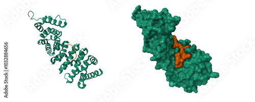
Adenomatous polyposis coli protein (green) with inhibitor (brown). 3D cartoon and molecular surface models, PDB 5z8h
PD-1 (violet) in complex with nivolumab Fab (brown and green). 3D cartoon and molecular surface model, entity id color scheme, PDB 5ggr
Wild type human transthyretin (TTR), a 3D ribbon model of the homodimer isolated, PDB 3w3b, white background
Structure of the main female hormone estradiol molecule, ball-and-stick model, white background, 3D illustration
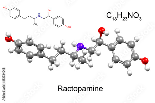
Chemical formula, structural formula and 3D ball-and-stick model of an animal feed additive ractopamine
Chemical formula, skeletal formula and 3D ball-and-stick model of a chemotherapeutic drug lenvatinib
Structure of the native human Angiotensin Converting Enzyme-Related Carboxypeptidase (ACE2), a receptor of SARS-CoV-2 spike glycoprotein, surface model, white background, 3D illustration
Crystal structure of Bacillus thuringiensis insecticidal crystal protein Cry7Ca1, 3D cartoon model, PDB 5zi1, secondary structure color scheme
Crystal structure of TAP01 in complex with amyloid beta peptide (green). 3D cartoon and molecular surface models, chain id color scheme, pdb 7ow1
Structure of glycosylated murine glutaminyl cyclase. 3D cartoon model, secondary structure color scheme, pdb 3si1
Cryo-EM structure of p62-PB1 filament (S-type). 3D cartoon and molecular surface models, chain id color scheme, pdb 6th3
Structure of human granulin A. 3D cartoon and molecular surface models, secondary structure color scheme, pdb 2jye
Crystal structure of TANK-binding kinase 1 (TBK1) with inhibitor bound. 3D cartoon and molecular surface models, chain id color scheme, pdb 4iwp
Structure of wild-type Troponin (C-I-T) complex. 3D cartoon and molecular surface models, chain id color scheme, pdb 8fmm
Complement component polyC9. 3D cartoon and molecular surface models, pdb 6dlw, putative membrane shown, chain id color scheme
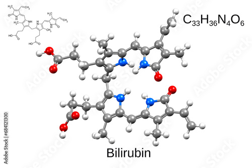
Chemical formula, skeletal formula, and 3D ball-and-stick model of bilirubin
Chemical formula, structural formula and 3D ball-and-stick model of an antifungal agent clotrimazole
Chemical formula, structural formula and 3D ball-and-stick model of antibiotic cefixime
Structure of human SRY-DNA complex. 3D cartoon and Gaussian surface models, PDB 1hrz
Crystal structure of the fat mass and obesity associated (FTO) protein. 3D cartoon model, secondary structure color scheme, PDB 3flm
Structure of human Sox2 (light brown) transcription factor in complex with a nucleosome. 3D cartoon model, secondary structure color scheme, PDB 6t7b
Structure of native human insulin oligomer. 3D cartoon and Gaussian surface models, chain id color scheme, PDB 4f1b
Cryo-EM structure of human kidney betaine-homocysteine methyltransferase. 3D cartoon and Gaussian surface models, chain id color scheme, PDB 8d45
Human alpha-galactosidase substarate bound. 3D cartoon and Gaussian surface models, PDB 3hg3, white background
Chemical formula, structural formula and 3D ball-and-stick model of a neurotransmitter serotonin, white background
Chemical formula, skeletal formula and 3D ball-and-stick model of the proton pump inhibitor, esomeprazole
Chemical formula, skeletal formula and 3D ball-and-stick model of a glutamate antagonist, riluzole
Chemical formula, structural formula and 3D ball-and-stick model of etoricoxib, a selective COX-2 inhibitor
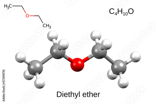
Chemical formula, skeletal formula and 3D ball-and-stick model of diethyl ether
Chemical formula, skeletal formula, and 3D ball-and-stick model of desflurane
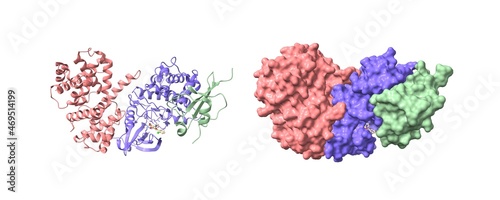
Crystal structure of trypsin (green)-vasopressin (brown) complex. 3D cartoon and Gaussian surface models, PDB 1yf4
Structure of insect steroid hormone 20-hydroxyecdysone. 3D ball-and-stick and space-filled models
DNA-binding domain of human SETMAR in complex with Hsmar1 terminal inverted repeat (TIR) DNA. 3D cartoon and Gaussian surface models, PDB 7s03
Structure of 6-aminohexanoate-oligomer hydrolase from Arthrobacter sp. KI72. 3D cartoon and Gaussian surface models, chain id color scheme, PDB 5y0m