Login
Sign Up
Member Area
Login
Sign Up
English
Afrikaans formal
Afrikaans informal
Arabic
Azerbaijan
Brazilian
Bulgarian
Catalan
Chinese simplified
Chinese traditional
Croatian
Czech
Danish
Dutch
English
Estonian
Finnish
French
Georgian
German
Greek
Hebrew
Hungarian
Icelandic
Indonesian
Italian
Japanese
Latvian
Lithuanian
Malaysian
Norwegian
Persian
Polish
Portuguese
Romanian
Russian
Serbian
Slovakian
Slovenian
Spanish
Swedish
Thai
Turkish
Ukrainian
Vietnamese
Toggle navigation
VideotonStock
Your Shopping Cart is empty.
Files
Stock
Photo
Video
Special offer
Collections
Packages
Lightboxes
Licenses
Royalty free
Rights managed
Creative
Editorial
Items
Most downloaded
Featured
Most popular
New
Free
Categories
Browse categories
Site info
Site info
About
Support
Privacy Policy
Privacy Policy
FAQ
Contact Us
Contact Us
Terms and Conditions
Buyer Agreement
Seller Agreement
Customers
Users
Credits
Photographers
Boby Richards
0
Boby Richards
0
Videoton1
286
Search
Keywords:
Creator ID:
Type:
Photo
Illustration
Vector
Video
Template
3d
Categories:
Animals
Buildings and Architecture
Business
Drinks
The Environment
States of Mind
Food
Graphic Resources
Hobbies and Leisure
Industry
Landscapes
Lifestyle
People
Plants and Flowers
Culture and Religion
Science
Social Issues
Sports
Technology
Transport
Travel
News
Entertainment
Sport News
Languages:
Brazilan Portuguese - Brazil
English - Canada
French - Canada
Spanish - Mexico
English - United States
English - Belgium
French - Belgium
Dutch - Belgium
Czech - Czech Republic
English - Cyprus
Danish - Danmark
German - Germany
English - Estonia
Spanish - Spain
French - France
French - Marocco
English - Greece
English - Ireland
Italian - Italia
English - Latvia
English - Lithuania
German - Luxembourg
English - Luxembourg
French - Luxembourg
English - Hungary
English - Malta
Dutch - Nederlands
Bokmal Norwegian - Norway
German - Austria
Polish - Poland
Portuguese - Portugal
English - Romania
German - Switzerland
English - Slovenia
Slovakian - Slovakia
French - Switzerland
English - Finland
Swedish- Sweden
Italian - Switzerland
English - United Kingdom
English - Bulgaria
English - Australia
Japanese - Japan
Korean - South Korea
English - New Zealand
Russian - Russia
English - Ukrain
English - Thailand
Orientation:
All
Horizontal
Vertical
square
Color:
Model property release:
All
Yes
No
Duration Video:
All
Up to 10 seconds
Up to 20 seconds
Longer than 30 seconds
Results
(0)
Site
Getty/iStock
Bigstockphoto
Pixabay
Adobe Stock
Relevance
New
Most popular
Most downloaded
Undiscovered
12
24
36
48
96
Auto paging
Menu
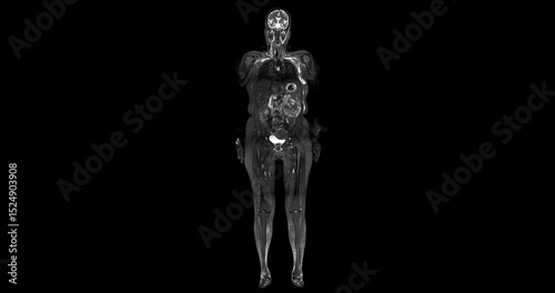
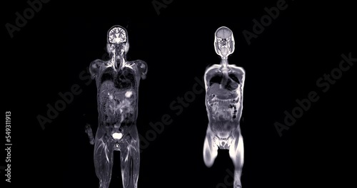
Full Body MRI Scan Visuals:An extraordinary journey through the body! This footage displays crisp, cross-sectional images of every organ and tissue from the brain to the limbs.
MRI prostate gland in aged mens for diagnosis prostate cancer cell .
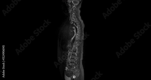
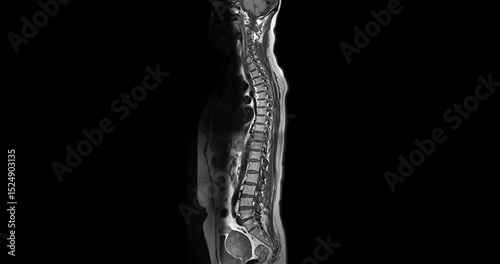
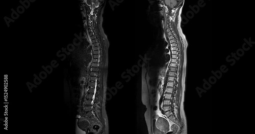
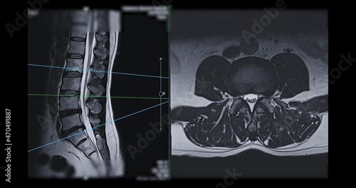
MRI Screening whole spine sagittal T2 FS showing spine compress spinal cord ( Myelopathy )
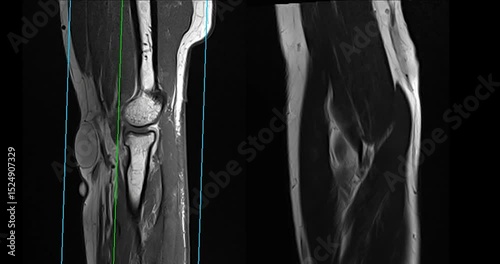
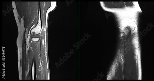
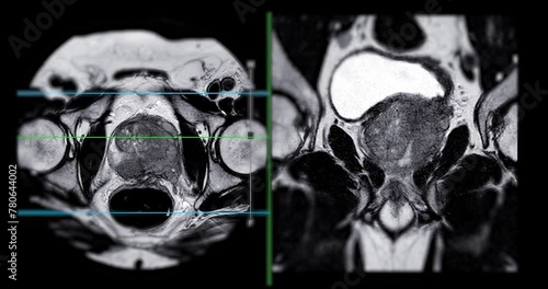
MRI elbow joint for Evaluation of the elbow by magnetic resonance imaging (MRI) is To facilitate accurate diagnosis.
MRI elbow joint for Evaluation of the elbow by magnetic resonance imaging (MRI) is To facilitate accurate diagnosis.
MRI Screening whole spine sagittal T2 FS showing spine compress spinal cord ( Myelopathy )
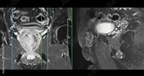
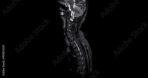
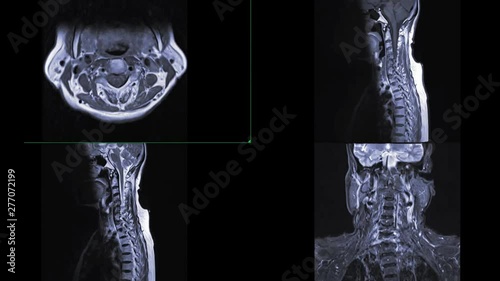
MRI C-spine or magnetic resonance image of cervical spine Coronal and sagittal T2W view for diagnosis spondylosis and compression fracture.
MRI C-spine or magnetic resonance image of cervical spine Coronal and sagittal T2W view for diagnosis spondylosis and compression fracture.
MRI elbow joint for Evaluation of the elbow by magnetic resonance imaging (MRI) is To facilitate accurate diagnosis.
MRI elbow joint for Evaluation of the elbow by magnetic resonance imaging (MRI) is To facilitate accurate diagnosis.
MRI elbow joint for Evaluation of the elbow by magnetic resonance imaging (MRI) is To facilitate accurate diagnosis.
MRI elbow joint for Evaluation of the elbow by magnetic resonance imaging (MRI) is To facilitate accurate diagnosis.
MRI whole body scans are beneficial for early cancer detection, monitoring chronic conditions, and overall health assessments.
MRI Screening whole spine sagittal T2 FS showing spine compress spinal cord ( Myelopathy )
MRI Screening whole spine sagittal T2 FS showing spine compress spinal cord ( Myelopathy )
MRI Screening whole spine sagittal T2 FS showing spine compress spinal cord ( Myelopathy )
MRI Screening whole spine sagittal T2 FS showing spine compress spinal cord ( Myelopathy )
MRI of the prostate gland, revealing an enlarged size, aids in diagnosing tumors, guiding treatment decisions, and monitoring prostate health.
Full Body MRI Scan Visuals:An extraordinary journey through the body! This footage displays crisp, cross-sectional images of every organ and tissue from the brain to the limbs.
MRI whole body scan Coronal T2 and T1 by MRI scanner.
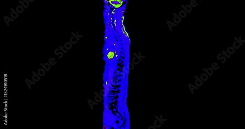
3D CTA of Lower Extremities Footage: A clear 3D visualization of the femoral artery and lower body skeleton, supporting vascular and orthopedic assessment.
High-resolution MRI breast footage displaying detailed soft tissue structures, aiding in the detection of tumors, cysts, and other abnormalities.
High-resolution MRI breast footage displaying detailed soft tissue structures, aiding in the detection of tumors, cysts, and other abnormalities.
MRI whole body scan Coronal T2 by MRI scanner.
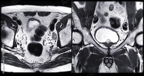
MRI L-S spine or lumbar spine Axial T2W view with sagittal plane for diagnosis spinal cord compression.
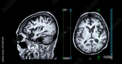
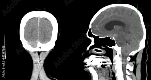
MRI of the brain compare sagittal and axial plane for diagnosis stroke diseases.
High-resolution MRI breast footage displaying detailed soft tissue structures, aiding in the detection of tumors, cysts, and other abnormalities.
Breast MRI magnetic resonance imaging of the breast uses radio waves and strong magnets to make detailed pictures of the inside of the breast To screen for breast cancer.
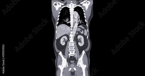
CT whole abdomen normal study: No abnormalities detected. All organs appear within normal limits with no signs of pathology.
Dynamic CTA brain scan showcasing detailed vascular structures, highlighting blood flow and arterial formations for medical diagnostics and research.
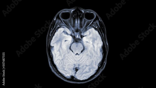
MRI of the brain axial t2 flair for detect brain stroke disease.
MRI of C-spine or magnetic resonance image of cervical spine sagittal view for diagnosis spondylosis and compression fracture.
A CT scan of the brain shows a subarachnoid hemorrhage scattered at both frontal lobes, temporal lobes, basal cistern and prepontine cistern., highlighting areas of bleeding.
MRI knee or Magnetic resonance imaging of knee joint for detect acl ligament tear.
MRI of the prostate gland reveals A 2.0x0.9x1.7 cm malignant prostate tumor at left posterior PZ, aiding in diagnosing tumors and guiding treatment decisions.
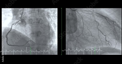
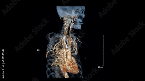
Cardiac catheterization showing coronary arteries used to diagnose and treat some heart conditions such as myocardial infarction.
MRI of the upper abdomen axial view is a non-invasive imaging technique providing detailed visuals of organs like the liver, pancreas, and kidneys.
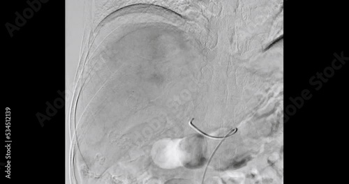
TOCE or Transcatheter Oily Chemo Embolization is a procedure that allows a dose of chemotherapy drugs to be administered directly to Liver tumor or HCC.
MRI knee or Magnetic resonance imaging of knee joint for detect acl ligament tear.
MRI of the brain sagittal plane for diagnosis stroke diseases.
A realistic 3D model of an CT scan machine in action with a patient undergoing the procedure. Ideal for educational and medical simulation purposes.
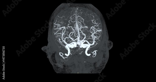
CTA Brain Vascular Footage highlights a detailed of the brain’s vascular network, showing clear visualization of Cerebral arteries and blood flow for diagnostic purposes.
CTA Brain Vascular Footage highlights a detailed of the brain’s vascular network, showing clear visualization of Cerebral arteries and blood flow for diagnostic purposes.
CTA Brain Vascular Footage highlights a detailed of the brain’s vascular network, showing clear visualization of Cerebral arteries and blood flow for diagnostic purposes.
CTA Brain Vascular Footage highlights a detailed of the brain’s vascular network, showing clear visualization of Cerebral arteries and blood flow for diagnostic purposes.
CTA Brain Vascular Footage highlights a detailed of the brain’s vascular network, showing clear visualization of Cerebral arteries and blood flow for diagnostic purposes.
CTA Brain Vascular Footage highlights a detailed of the brain’s vascular network, showing clear visualization of Cerebral arteries and blood flow for diagnostic purposes.
CTA Brain Vascular Footage highlights a detailed of the brain’s vascular network, showing clear visualization of Cerebral arteries and blood flow for diagnostic purposes.
CTA Brain Vascular Footage highlights a detailed of the brain’s vascular network, showing clear visualization of Cerebral arteries and blood flow for diagnostic purposes.
CTA Brain Vascular Footage highlights a detailed of the brain’s vascular network, showing clear visualization of Cerebral arteries and blood flow for diagnostic purposes.
CTA Brain Vascular Footage highlights a detailed of the brain’s vascular network, showing clear visualization of Cerebral arteries and blood flow for diagnostic purposes.
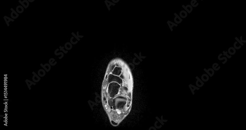
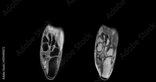
MRI foot scan for a diagnostic of the foot and ankle following symptoms: pain both feet, swelling, redness or warmth, numbness or tingling and stiffness.
MRI foot scan for a diagnostic of the foot and ankle following symptoms: pain both feet, swelling, redness or warmth, numbness or tingling and stiffness.
MRI foot scan for a diagnostic of the foot and ankle following symptoms: pain both feet, swelling, redness or warmth, numbness or tingling and stiffness.
MRI foot scan for a diagnostic of the foot and ankle following symptoms: pain both feet, swelling, redness or warmth, numbness or tingling and stiffness.
MRI foot scan for a diagnostic of the foot and ankle following symptoms: pain both feet, swelling, redness or warmth, numbness or tingling and stiffness.
MRI foot scan for a diagnostic of the foot and ankle following symptoms: pain both feet, swelling, redness or warmth, numbness or tingling and stiffness.
CT Scan of Knee joint.
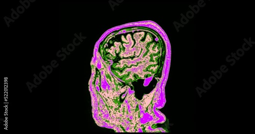
The MRI brainT2 flair sequence scan footage reveals intricate brain details, aiding in diagnostic procedures.
Femoral Angiogram of femoral artery showing femoral artery vessel isolated on white background for diagnosis atherosclerosis.
CT Scan of chest and abdomen in colorful mode or Pet scan.
MRI of the prostate gland reveals a focal abnormal signal intensity (SI) lesion at the left posterolateral peripheral zones at the apex, aiding in diagnosing tumors and guiding treatment decisions.
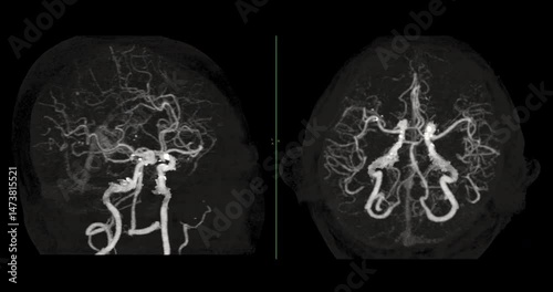
Comparison of MRI MRA brain for detect Brain disease.
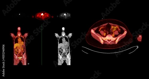
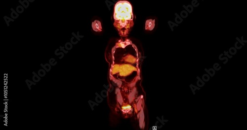
A PET-CT scan is a diagnostic imaging technique combining positron emission tomography (PET) and computed tomography (CT) in cancer diagnosis and staging.
CT Scan L-S pine Showing T12 Compression Injury, where the front part of the bone has collapsed slightly. The fracture looks stable, and there is no damage to the spinal cord or nearby tissues.
The MRI brain footage reveals areas of reduced blood flow, indicating early signs of stroke disease.
CTA thoracic aorta include common carotid artery 3D rendering with perm catheter on bone transparency image in case Dialysis patients
CT whole abdomen normal study: No abnormalities detected. All organs appear within normal limits with no signs of pathology.
CTA Whole Aorta with Stent-Grafts Coronal view: CTA scan of the whole aorta, highlighting thoracic aorta endovascular stent-grafts placed for aneurysm repair and blood flow restoration.
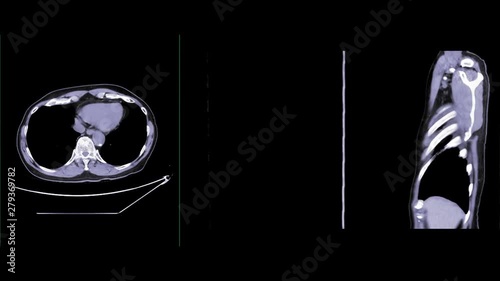
CT Chest Sagittal view Normal Study: CT chest footage reveals normal lung, heart, and mediastinal structures, showing no signs of abnormalities, ensuring a healthy thoracic region.
CT Chest Axial view Normal Study: CT chest footage reveals normal lung, heart, and mediastinal structures, showing no signs of abnormalities, ensuring a healthy thoracic region.
CT Chest with Contrast or CTA pulmonary artery axial NC ,Coronal and sagittal view for diagnosis pulmonary embolism and lung disease.
Comparison of MRI C-spine or magnetic resonance image of cervical spine Axial , Coronal and sagittal plane showing spondylosis and compression fracture.
The MRI brain footage reveals areas of reduced blood flow, indicating early signs of stroke disease.
The MRI brain footage reveals areas of reduced blood flow, indicating early signs of stroke disease.
A PET-CT scan is a diagnostic imaging technique combining positron emission tomography (PET) and computed tomography (CT) in cancer diagnosis and staging.
A PET-CT scan is a diagnostic imaging technique combining positron emission tomography (PET) and computed tomography (CT) in cancer diagnosis and staging.
A PET-CT scan is a diagnostic imaging technique combining positron emission tomography (PET) and computed tomography (CT) in cancer diagnosis and staging.
A PET-CT scan is a diagnostic imaging technique combining positron emission tomography (PET) and computed tomography (CT) in cancer diagnosis and staging.
CT Chest or CT Scan of Lung View for diagnosis TB,tuberculosis and coronavirus or covid-19 .
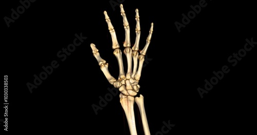
CT scan footage showing a detailed 3D rendering of a normal human hand. The scan highlights clear bone structures, joints, and soft tissues.
The MRI brain footage reveals areas of reduced blood flow, indicating early signs of stroke disease.
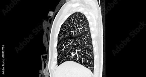
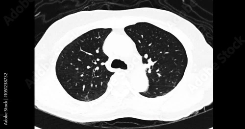
The CT scan footage shows clear and well-defined lung tissues, indicating a normal case with no signs of abnormalities.
The CT scan footage shows clear and well-defined lung tissues, indicating a normal case with no signs of abnormalities.
Footage of an MRI orbit scan in normal case displays comprehensive views for eye disease diagnosis.
A PET CT scan reveals cholangiocarcinoma with suspected metastatic involvement of the lungs and lymph nodes.
A CT scan of the brain shows a subarachnoid hemorrhage scattered at both frontal lobes, temporal lobes, basal cistern and prepontine cistern., highlighting areas of bleeding.
A CT scan of the brain shows a subarachnoid hemorrhage scattered at both frontal lobes, temporal lobes, basal cistern and prepontine cistern., highlighting areas of bleeding.
CT Abdomen show a retroperitoneal mass with a peripheral fatty component with thin septa and poorly defined solid-appearing areas with no clear demarcation between them and fat.
computed tomography ,CT scan of thoracic spine 2D and 3D rendering image . Medical technology concept.
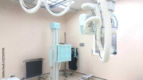
X-ray generator tube / X-ray machine moving on the position automatic control. modern medical equipment.
MRI of the prostate gland, revealing an enlarged size, aids in diagnosing tumors, guiding treatment decisions, and monitoring prostate health.
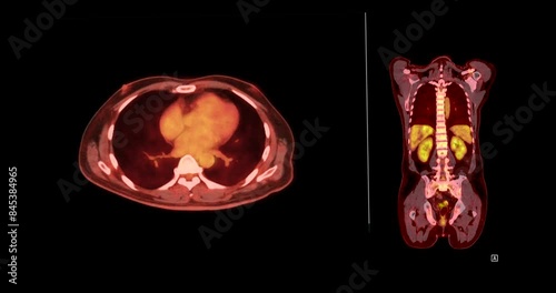
PET CT Scan fusion image of Whole human body for detect cancer recurrence after surgery .
CT scan of the brain for diagnosis brain tumor,stroke diseases and vascular diseases.
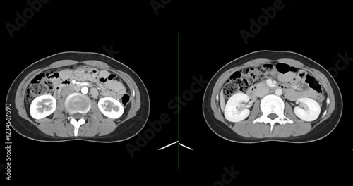
A detailed CT scan showing the human kidney and vascular system, offering a close-up look at the anatomy and structure of these vital organs.
A detailed CT scan showing the human kidney and vascular system, offering a close-up look at the anatomy and structure of these vital organs.
A detailed CT scan showing the human kidney and vascular system, offering a close-up look at the anatomy and structure of these vital organs.
Breast MRI revealing BI-RADS 4 in women indicates suspicious findings warranting further investigation for potential malignancy and biopsy to confirm the presence of cancerous lesions.
A PET-CT scan is a diagnostic imaging technique combining positron emission tomography (PET) and computed tomography (CT) in cancer diagnosis and staging.
MRI scan reveals a lesion in the left hemisphere of the brain, providing critical insight into the patient's condition for diagnosis and treatment planning.