Login
Sign Up
Member Area
Login
Sign Up
English
Afrikaans formal
Afrikaans informal
Arabic
Azerbaijan
Brazilian
Bulgarian
Catalan
Chinese simplified
Chinese traditional
Croatian
Czech
Danish
Dutch
English
Estonian
Finnish
French
Georgian
German
Greek
Hebrew
Hungarian
Icelandic
Indonesian
Italian
Japanese
Latvian
Lithuanian
Malaysian
Norwegian
Persian
Polish
Portuguese
Romanian
Russian
Serbian
Slovakian
Slovenian
Spanish
Swedish
Thai
Turkish
Ukrainian
Vietnamese
Toggle navigation
VideotonStock
Your Shopping Cart is empty.
Files
Stock
Photo
Video
Special offer
Collections
Packages
Lightboxes
Licenses
Royalty free
Rights managed
Creative
Editorial
Items
Most downloaded
Featured
Most popular
New
Free
Categories
Browse categories
Site info
Site info
About
Support
Privacy Policy
Privacy Policy
FAQ
Contact Us
Contact Us
Terms and Conditions
Buyer Agreement
Seller Agreement
Customers
Users
Credits
Photographers
Boby Richards
0
Boby Richards
0
Videoton1
286
Search
Keywords:
Creator ID:
Type:
Photo
Illustration
Vector
Video
Template
3d
Categories:
Animals
Buildings and Architecture
Business
Drinks
The Environment
States of Mind
Food
Graphic Resources
Hobbies and Leisure
Industry
Landscapes
Lifestyle
People
Plants and Flowers
Culture and Religion
Science
Social Issues
Sports
Technology
Transport
Travel
News
Entertainment
Sport News
Languages:
Brazilan Portuguese - Brazil
English - Canada
French - Canada
Spanish - Mexico
English - United States
English - Belgium
French - Belgium
Dutch - Belgium
Czech - Czech Republic
English - Cyprus
Danish - Danmark
German - Germany
English - Estonia
Spanish - Spain
French - France
French - Marocco
English - Greece
English - Ireland
Italian - Italia
English - Latvia
English - Lithuania
German - Luxembourg
English - Luxembourg
French - Luxembourg
English - Hungary
English - Malta
Dutch - Nederlands
Bokmal Norwegian - Norway
German - Austria
Polish - Poland
Portuguese - Portugal
English - Romania
German - Switzerland
English - Slovenia
Slovakian - Slovakia
French - Switzerland
English - Finland
Swedish- Sweden
Italian - Switzerland
English - United Kingdom
English - Bulgaria
English - Australia
Japanese - Japan
Korean - South Korea
English - New Zealand
Russian - Russia
English - Ukrain
English - Thailand
Orientation:
All
Horizontal
Vertical
square
Color:
Model property release:
All
Yes
No
Duration Video:
All
Up to 10 seconds
Up to 20 seconds
Longer than 30 seconds
Results
(0)
Site
Getty/iStock
Bigstockphoto
Pixabay
Adobe Stock
Relevance
New
Most popular
Most downloaded
Undiscovered
12
24
36
48
96
Auto paging
Menu
ECG Atrial Fibrillation - 8 Second ECG Paper - Electrocardiography Vector Medical Illustration
Conducting System of the Heart - Medical Vector Illustration
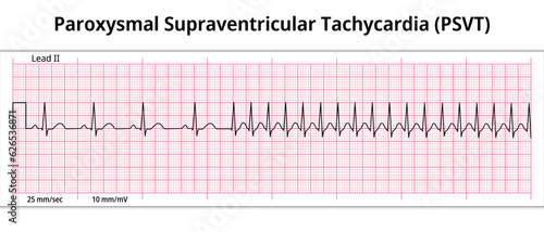
ECG Paroxysmal Supraventricular Tachycardia (PSVT) - 8 Second ECG Paper - Electrocardiogram Vector Medical Illustration
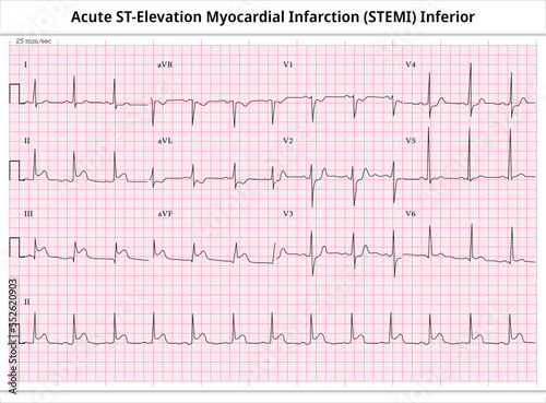
Acute ST-Elevation Myocardial Infarction (STEMI) Inferior - ECG Paper 12 Lead
Normal Range ECG - PR Interval, QRS Complex, QT Interval, P and T Wave - Electrocardiogram Medical Vector Illustration
Set of ECG Common Abnormalities - Atrial Flutter vs Atrial Fibrillation - Normal Sinus Rhythm - Electrocardiography Vector Medical Illustration
ECG Junctional Rhythm - Escape Rhythm - 8 Second ECG Paper - Electrocardiogram Vector Medical Illustration
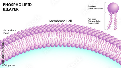
Structure of the Phospholipid Bilayer in the Cell Membrane - 3D look Medical Vector Illustration
ECG Sinus Arrhythmia - 8 Second ECG Paper - Electrocardiogram Vector Medical Illustration
Standard ECG Paper - grid of squares that moves at a speed of 25 mm/s and a voltage of 10 mm/mV - Electrocardiogram Medical Vector Illustration
ECG Ventricular Tachycardia – Monomorphic VT - 8 Second ECG Paper
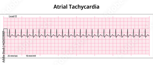
ECG Atrial Tachycardia - 8 Second ECG Paper - Electrocardiography Vector Medical Illustration
Set of ECG Common Abnormalities - Normal Sinus Rhythm - Sinus Tachycardia - Sinus Bradycardia - Vector Medical Illustration
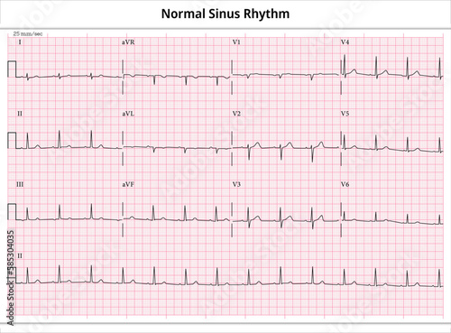
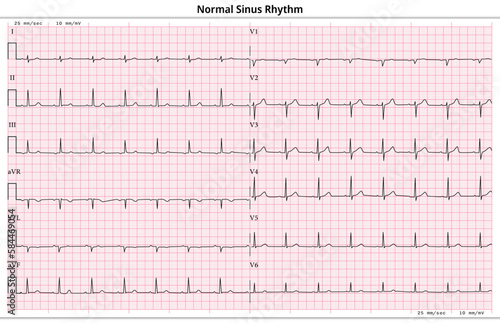
ECG Normal Sinus Rhythm - 12 Lead ECG Common Case - 3 Sec/lead - Vector Illustration
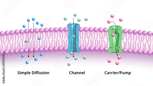
Major Mechanisms by which Molecules Cross the Cell Membrane, Simple Diffusion, Channel, and Pump / Carrier - Medical Vector Illustration
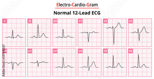
The Differences in Normal ECG Waveform for Each of the 12 Leads Standard ECG - Medical Vectors and Illustrations for Medical Purposes
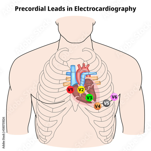
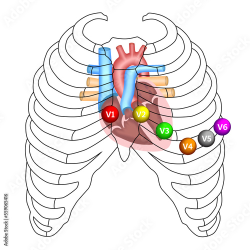
Precordial Leads in Electrocardiography - V1, V2, V3, V4, V5, and V6 - Position of ECG Chest Leads - Vector Medical Illustration
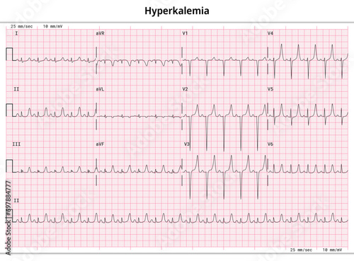
ECG Hyperkalemia - Hyperkalaemia - 12 Lead ECG Common Case - 3 Sec/lead - Vector Medical Illustration
ECG Sinus Tachycardia - 8 Second ECG Paper
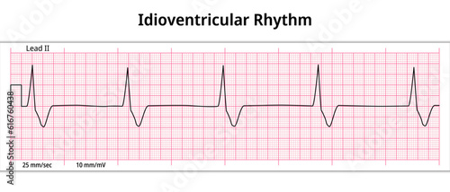
ECG Idioventricular Rhythm - 8 Second ECG Paper - Electrocardiography Medical Vector Illustration
General Structure of a Phospholipid - Structural Formula - Vector Medical Illustration
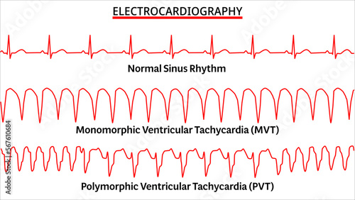
Set of ECG Common Abnormalities - Normal Sinus Rhythm - Monomorphic VT - Polymorphic VT - Vector Medical Illustration
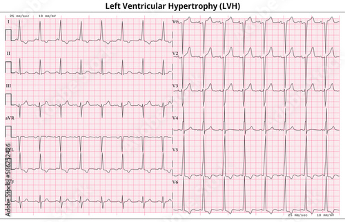
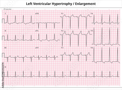
ECG Left Ventricular Hypertrophy (LVH) - Left Ventricular Enlargement - 12 Lead ECG Common Case - 6 Sec/lead - Vector Illustration
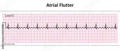
ECG Atrial Flutter - 8 Second ECG Paper
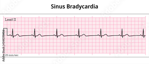
ECG Sinus Bradycardia - 8 Second ECG Paper
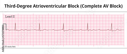
ECG Third Degree Atrioventricular Block - Complete AV Block - 8 Second ECG Paper - Electrocardiography Vector Medical Illustration
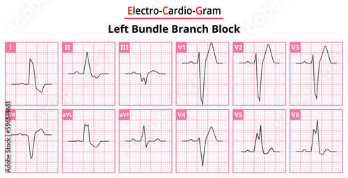
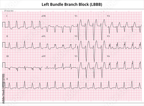
Left Bundle Branch Block - The Differences in ECG Waveform for Each of the 12 Leads - Medical Vector and Illustration
ECG Ventricular Pacemaker Rhythm - Single Chamber - 8 Second ECG Paper - Electrocardiography Vector Medical Illustration
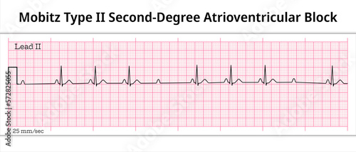
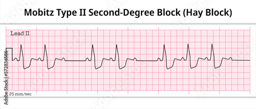
ECG Mobitz Type 2 Second Degree Atrioventricular Block - 2nd degree AV Block - 8 Second ECG Paper - Electrocardiography Vector Medical Illustration
ECG Normal Sinus Rhythm - 12 Lead ECG Common Case - 6 Sec/lead - Vector Medical Illustration
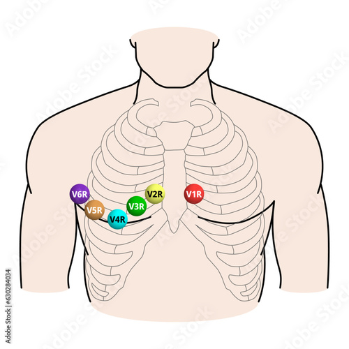
Right Sided ECG Electrode Placement - Electrocardiogram Lead V1R, V2R, V3R, V4R, V5R, V6R Positions - Medical Vector Illustration
ECG Asystole - 8 Second ECG Paper
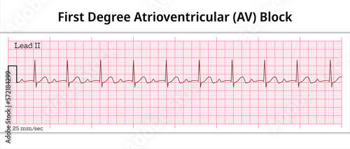
ECG First Degree Atrioventricular Block - 1st degree AV Block - 8 Second ECG Paper - Electrocardiography Vector Medical Illustration
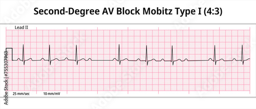
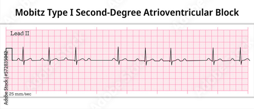
ECG Second Degree AV Block Mobitz Type 1 (4:3) - 8 Second Paper Electrocardiogram - Medical Vector Illustration
Extensive Anterolateral STEMI (ST-Elevation Myocardial Infarction) Acute - ECG Paper 12 Lead - Vector Medical Illustration
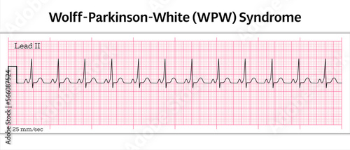
ECG Wolff-Parkinson-White (WPW) Syndrome - 8 Second ECG Paper - Vector Medical Illustration
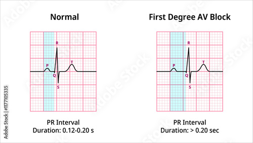
Comparison of AV Block Degree 1 and Normal ECG Waveforms - Medical Vector Illustration
Fine Atrial Fibrillation - 8 Second ECG Paper - Electrocardiography Vector Medical Illustration
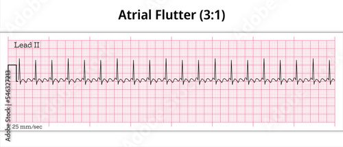
ECG Atrial Flutter (3:1)- 8 Second ECG Paper
ECG P wave Asystole - Ventricular Standstill - 8 Second ECG Paper - Electrocardiogram Medical Vector Illustration
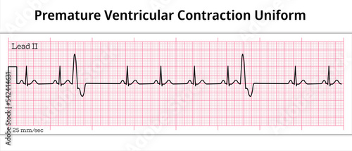
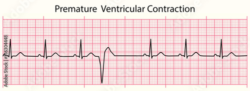
ECG Premature Ventricular Contraction Uniform - Unifocal - 8 Second ECG Paper - Ventricular Extrasystole
ECG Second Degree AV Block Mobitz Type 2 (2:1) - 8 Sec Electrocardiogram - Medical Vector Illustration
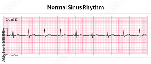
ECG Normal Sinus Rhythm - 8 Second ECG Paper - Vector Medical Illustration
ECG Wolff-Parkinson-White (WPW) Syndrome - 8 Second ECG Paper - Vector Medical Illustration
ECG Pulseless Electrical Activity (PEA) - 8 Second ECG Paper - Electrocardiogram Vector Medical Illustration
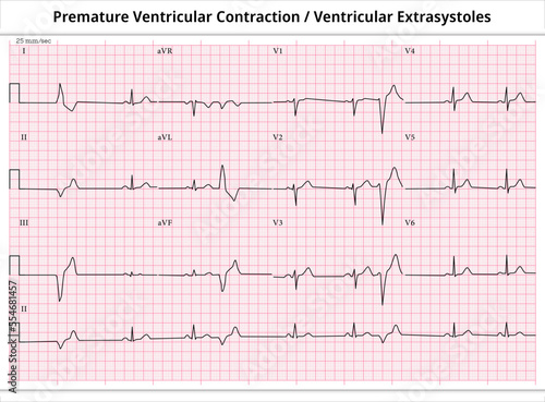
ECG PVC (Premature Ventricular Contractions) - ECG VES (Ventricular Extrasystoles) - 12 Leads ECG Paper
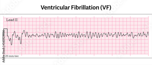
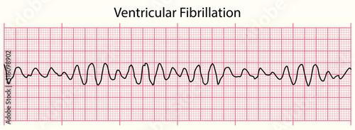
ECG Ventricular Fibrillation (VF) - 8 Second ECG Paper - Vector Medical Illustration
Phospholipid Bilayer Structure - Single Layer - Medical Vector Illustration.
ECG Precordial Lead Position - V1, V2, V3, V4, V5, and V6 - Position of ECG chest leads - Vector Medical Illustration
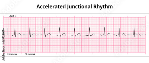
ECG Accelerated Junctional Rhythm - Escape Rhythm - 8 Second ECG Paper - Electrocardiogram Vector Medical Illustration
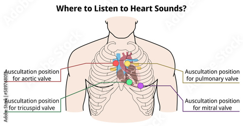
Where to listen to heart sounds? - Auscultation Position of Heart Valve - Vector Medical Illustration
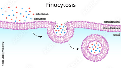
Pinocytosis - Process by which The Cell Takes in The Fluids Along with Dissolved Small Molecules - Medical Vector Illustration
Wenckebach Phenomenon - ECG Mobitz Type 1 Second Degree Atrioventricular Block - 2nd degree AV Block - 8 Second ECG Paper - Electrocardiography Vector Medical Illustration
ECG Torsade de Pointes - Long QT Interval - Ventricular Tachycardia - 12 Leads ECG Paper - Vector Medical Illustration
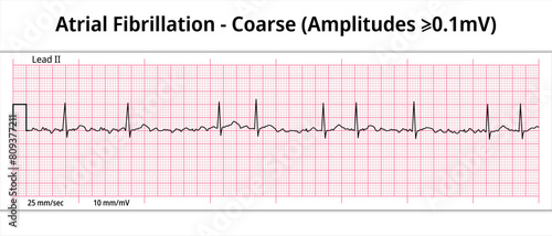
Coarse Atrial Fibrillation - 8 Second ECG Paper - Electrocardiography Vector Medical Illustration
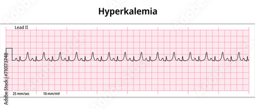
ECG Hyperkalemia - Hyperkalaemia - 8 Second ECG Paper - Electrocardiogram Vector Medical Illustration
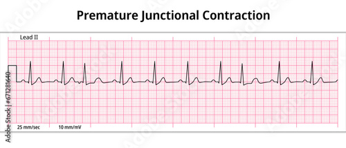
ECG Premature Junctional Contraction - 8 Second ECG Paper - Electrocardiogram Vector Medical Illustration
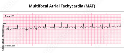
Multifocal Atrial Tachycardia (MAT) - 8 Second ECG Paper - Electrocardiography Vector Medical Illustration
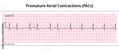
Premature Atrial Contractions (PACs) - Premature Atrial Complex (PAC) - 8 Second ECG Paper - Electrocardiography Vector Medical Illustration
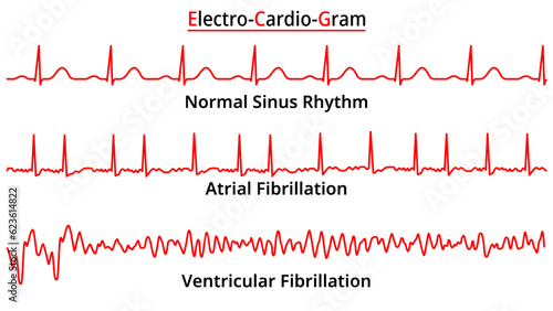
Set of ECG Common Abnormalities - Atrial Fibrillation - Ventricular Fibrillation - Normal Sinus Rhythm - Electrocardiography Vector Medical Illustration
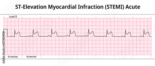
ST-Elevation Myocardial Infarction (STEMI) Acute - 8 Second ECG Paper - Electrocardiography Vector Medical Illustration
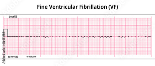
ECG Fine Ventricular Fibrillation - 8 Second ECG Paper - Electrocardiography Medical Vector Illustration
ECG line: Premature Ventricular Contraction in 6 second ECG paper line
ECG Torsades de Pointes Ventricular Tachycardia - 8 Second ECG Paper
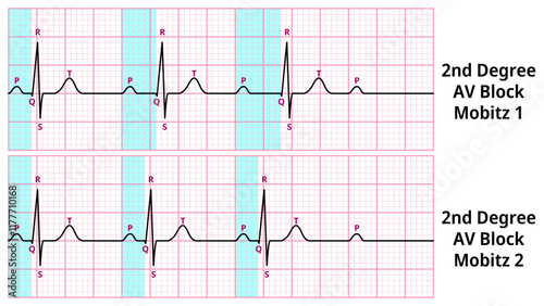
Comparison of AV Block Degree 2 Mobitz Type 1 and Type 2 ECG Waveforms - Medical Vector Illustration
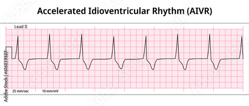
ECG Accelerated Idioventricular Rhythm - 8 Second ECG Paper - Electrocardiography Medical Vector Illustration
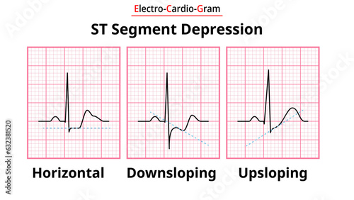
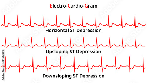
ECG Morphology of ST Segment Depression - Horizontal, Upsloping, and Downsloping - Medical Electrocardiogram Vector Illustration
ECG Torsades de Pointes Ventricular Tachycardia - 8 Second ECG Paper
ECG Atrial Ventricular Pacemaker Rhythm - Dual Chamber - 8 Second ECG Paper - Electrocardiography Vector Medical Illustration
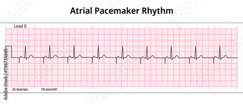
ECG Atrial Pacemaker Rhythm - Single Chamber - 8 Second ECG Paper - Electrocardiogram Vector Medical Illustration
ECG Hypokalemia - Hypokalaemia - 12 Lead ECG Common Case - 3 Sec/lead - Electrocardiogram Vector Medical Illustration
Receptor Mediated Endocytosis - RME - Clathrin Mediated Endocytosis - Process by which cells absorb metabolites, hormones, proteins, virus - Medical Vector Illustration
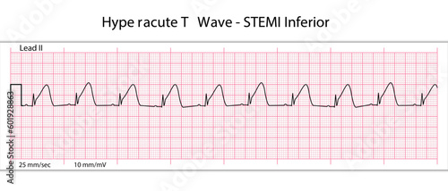
ECG Hyperacute STEMI - 8 Second ECG Paper - Peaked T Wave - Vectors and Illustrations for Medical Purposes
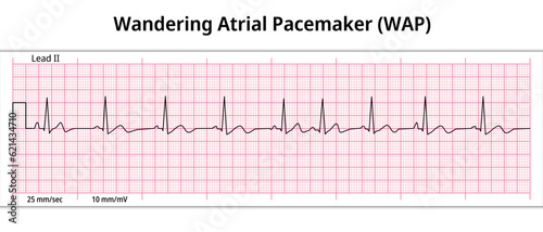
ECG Wandering Atrial Pacemaker (WAP) - 8 Second ECG Paper - Electrocardiography Vector Medical Illustration
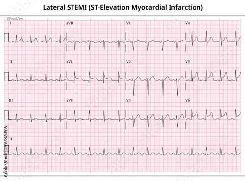
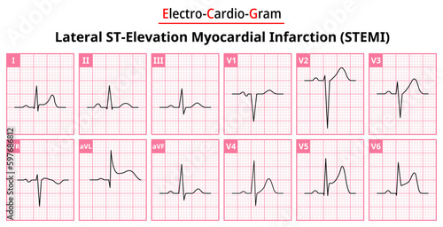
Lateral ST-Elevation Myocardial Infarction (STEMI) - 12 Lead ECG Common Case - 3 Sec/lead - Vectors and Illustration for Medical Purposes
ECG Right Atrial Enlargement (RAE) - Right Atrial Hypertrophy (RAH) - 12 Lead ECG Common Case - 6 Sec/lead - Electrocardiogram Medical Vector Illustration
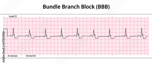
ECG Bundle Branch Block - 8 Second ECG Paper - Electrocardiogram Vector Medical Illustration
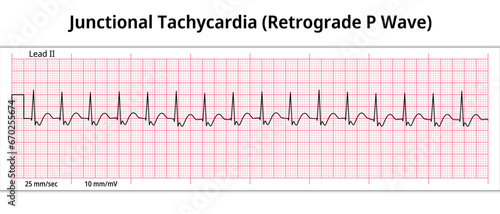
ECG Junctional Tachycardia - Retrograde P Wave - 8 Second ECG Paper - Electrocardiogram Vector Medical Illustration
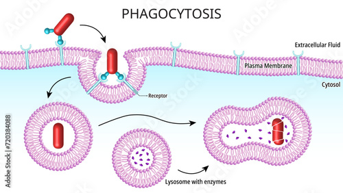
Phagocytosis - Process for nutrition in unicellular organisms, while in multicellular organisms it is found in specialized cells called phagocytes - Cell Medical Vector Illustration
ECG Monomorphic VT - Ventricular Tachycardia - 12 Leads ECG Paper - Vectors
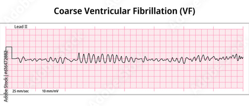
ECG Coarse Ventricular Fibrillation - 8 Second ECG Paper - Electrocardiography Medical Vector Illustration
ECG line: Ventricular Fibrillation in 6 second ECG paper line
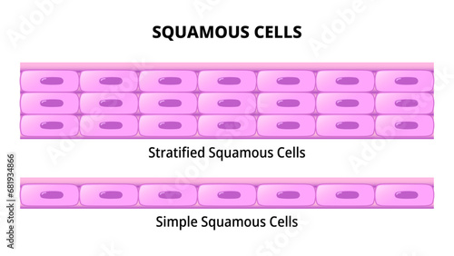
Squamous Cell - Simple Squamous Epithelium - Stratified Squamous Epithelium - Histology Medical Vector Illustration
ECG Acute STEMI (ST-Elevation Myocardial Infarction) - 12 Lead ECG Common Case - 6 Sec/lead - Vector Illustration
Facilitated Diffusion - Membrane Transport - Molecules Across Cell Membrane - Medical Vector Illustration
Bigeminy Premature Ventricular Contractions (PVCs) - Ventricular Premature Beat - VES - 8 Second ECG Paper - Electrocardiography Vector Medical Illustration
ECG Mobitz Type 1 Second Degree Atrioventricular Block - 2nd degree AV Block - 8 Second ECG Paper - Electrocardiography Vector Medical Illustration
Hay Block - ECG Mobitz Type 2 Second Degree Atrioventricular Block - 2nd degree AV Block - 8 Second ECG Paper - Electrocardiography Vector Medical Illustration
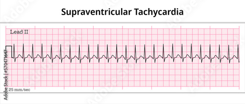
ECG Supraventricular Tachycardia - 8 Second ECG Paper - Electrocardiography Vector Medical Illustration
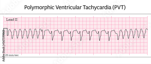
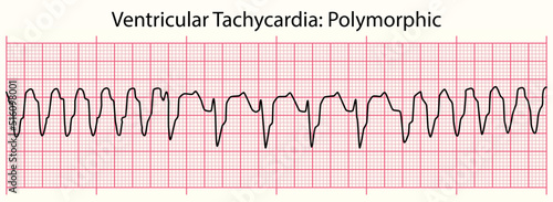
Polymorphic Ventricular Tachycardia (PVT) ECG – Polymorphic VT - 8 Second ECG Paper - Vector Medical Illustration
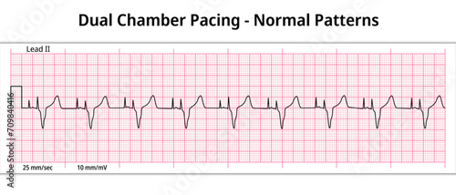
ECG Dual Chamber Pacemaker Rhythm - Normal Pattern - 8 Second ECG Paper - Electrocardiography Vector Medical Illustration
ECG Left Bundle Branch Block - ECG Paper 12 Lead
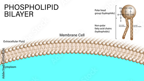
PHOSPHOLIPID BILAYER - MEMBRANE CELL
Set of ECG Common Abnormalities - ST Segment Depression - Upsloping - Downsloping - Horizontal - Electrocardiogram Vector Medical Illustration
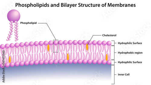
Phospholipids and Bilayer Structure of Membranes - Vector Medical Illustration
ST-Elevation Myocardial Infarction (STEMI) Sub-Acute - 8 Second ECG Paper - Electrocardiography Vector Medical Illustration
ECG Left Ventricular Enlargement - ECG Paper 12 Leads - Left Ventricular Hypertrophy
ECG line: Ventricular Tachycardia Polymorphic in 6 second ECG paper line
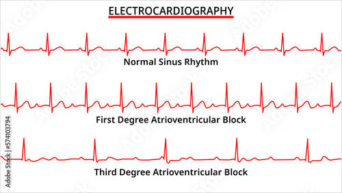
Set of ECG Common Abnormalities - Normal Sinus Rhythm - First and Third Degree Atrioventricular (AV) Block - Electrocardiography Vector Medical Illustration
Lateral STEMI (ST-Elevation Myocardial Infarction) - The Differences in ECG Waveform for Each of the 12 Leads - Vectors and Illustrations for Medical Purposes