Login
Sign Up
Member Area
Login
Sign Up
English
Afrikaans formal
Afrikaans informal
Arabic
Azerbaijan
Brazilian
Bulgarian
Catalan
Chinese simplified
Chinese traditional
Croatian
Czech
Danish
Dutch
English
Estonian
Finnish
French
Georgian
German
Greek
Hebrew
Hungarian
Icelandic
Indonesian
Italian
Japanese
Latvian
Lithuanian
Malaysian
Norwegian
Persian
Polish
Portuguese
Romanian
Russian
Serbian
Slovakian
Slovenian
Spanish
Swedish
Thai
Turkish
Ukrainian
Vietnamese
Toggle navigation
VideotonStock
Your Shopping Cart is empty.
Files
Stock
Photo
Video
Special offer
Collections
Packages
Lightboxes
Licenses
Royalty free
Rights managed
Creative
Editorial
Items
Most downloaded
Featured
Most popular
New
Free
Categories
Browse categories
Site info
Site info
About
Support
Privacy Policy
Privacy Policy
FAQ
Contact Us
Contact Us
Terms and Conditions
Buyer Agreement
Seller Agreement
Customers
Users
Credits
Photographers
Boby Richards
0
Boby Richards
0
Videoton1
286
Search
Keywords:
Creator ID:
Type:
Photo
Illustration
Vector
Video
Template
3d
Categories:
Animals
Buildings and Architecture
Business
Drinks
The Environment
States of Mind
Food
Graphic Resources
Hobbies and Leisure
Industry
Landscapes
Lifestyle
People
Plants and Flowers
Culture and Religion
Science
Social Issues
Sports
Technology
Transport
Travel
News
Entertainment
Sport News
Languages:
Brazilan Portuguese - Brazil
English - Canada
French - Canada
Spanish - Mexico
English - United States
English - Belgium
French - Belgium
Dutch - Belgium
Czech - Czech Republic
English - Cyprus
Danish - Danmark
German - Germany
English - Estonia
Spanish - Spain
French - France
French - Marocco
English - Greece
English - Ireland
Italian - Italia
English - Latvia
English - Lithuania
German - Luxembourg
English - Luxembourg
French - Luxembourg
English - Hungary
English - Malta
Dutch - Nederlands
Bokmal Norwegian - Norway
German - Austria
Polish - Poland
Portuguese - Portugal
English - Romania
German - Switzerland
English - Slovenia
Slovakian - Slovakia
French - Switzerland
English - Finland
Swedish- Sweden
Italian - Switzerland
English - United Kingdom
English - Bulgaria
English - Australia
Japanese - Japan
Korean - South Korea
English - New Zealand
Russian - Russia
English - Ukrain
English - Thailand
Orientation:
All
Horizontal
Vertical
square
Color:
Model property release:
All
Yes
No
Duration Video:
All
Up to 10 seconds
Up to 20 seconds
Longer than 30 seconds
Results
(0)
Site
Getty/iStock
Bigstockphoto
Pixabay
Adobe Stock
Relevance
New
Most popular
Most downloaded
Undiscovered
12
24
36
48
96
Auto paging
Menu
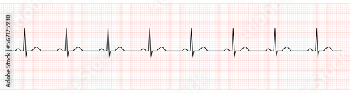
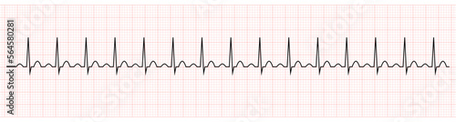
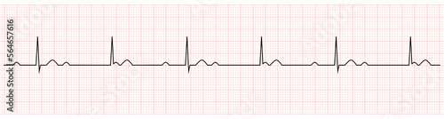
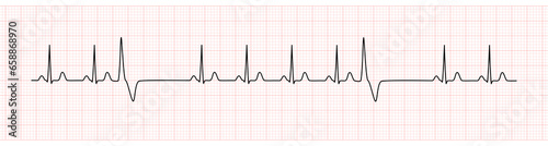
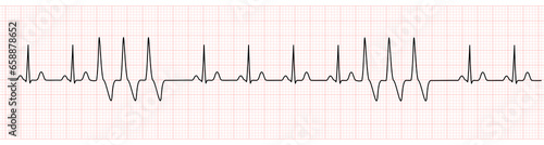
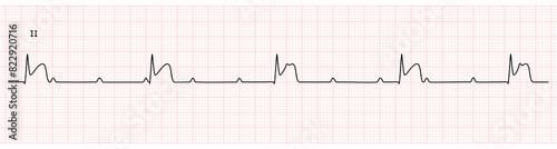
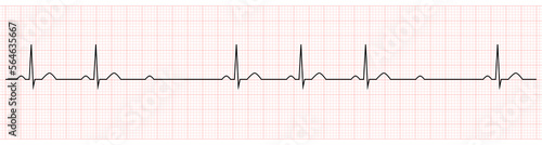
EKG showing normal sinus rhythm of patient
EKG Monitor in lead II Showing Sinus Rhythm with Late STEMI at Inferior Wall with Deep Q Wave
EKG Monitor Showing Sinus Rhythm with Short QT interval
EKG Monitor Showing Sinus Rhythm with prolong QT interval
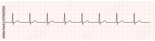
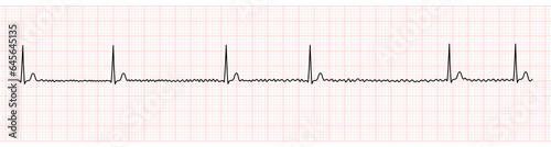
EKG Monitor Showing junctional bradycardia
EKG Monitor Showing Polymorphic Ventricular tachycardia or VT: Torsades de pointes
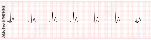
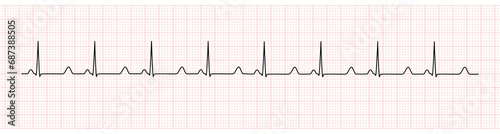
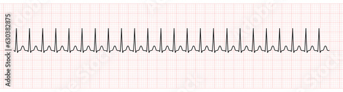
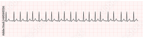
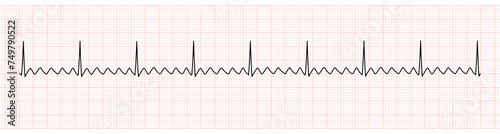
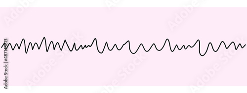
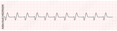
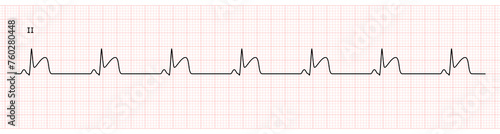
EKG Showing Sinus tachycardia of Patient
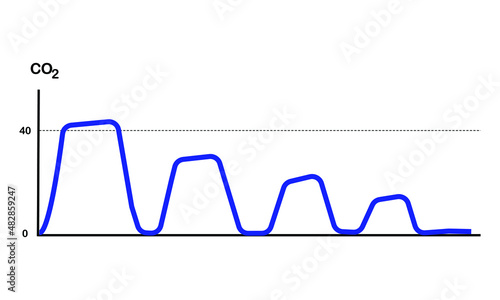
capnography waveform during high quality CPR
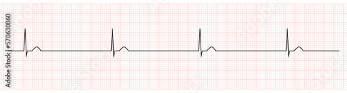
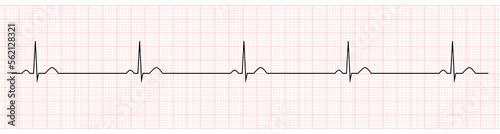
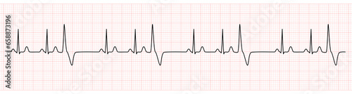
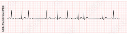
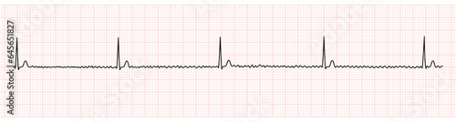
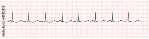
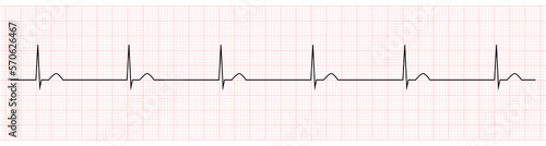
EKG Showing Sinus Bradycardia of Patient
EKG Showing third degree or complete av block
capnography waveform of endotracheal tube displacement from airway
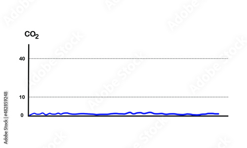
capnography waveform of esophageal intubation
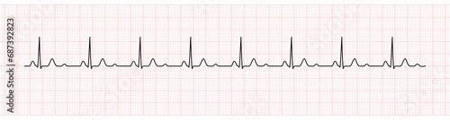
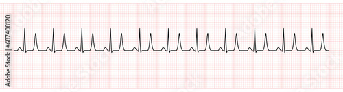
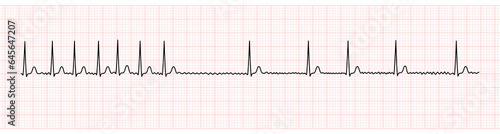
EKG Monitor Showing Supraventricular Tachycardia
Icon For Airway Emergency Management Equipment. Healthcare and Medical Concept
normal oxygen saturation waveform with poor baseline waveform monitoring
EKG Showing Sinus Bradycardia with Arrest of Patient
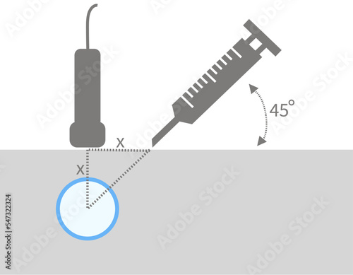
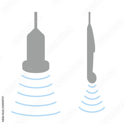
Animation of Method of Ultrasound Guided Central Venous Catheter Insertion
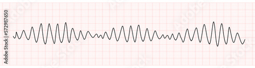
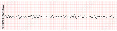
EKG Monitor Showing Ventricular Fibrillation or VF
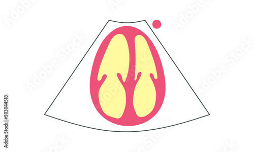
Infographic of Echocardiogram of Apical Five Chamber And Measurement of LVOT VTI
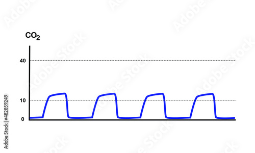
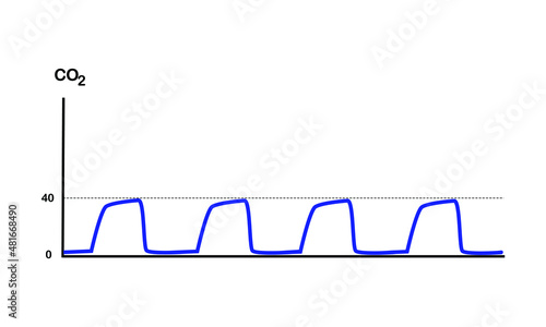
picture of Normal Capnography on white background
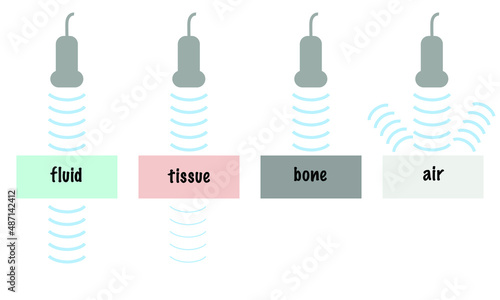
Ultrasound Wave Attenuation of Different Tissue Including Fluid Soft Tissue Bone Air
Diagram of Inferior Vena Cava Ultrasound in M Mode
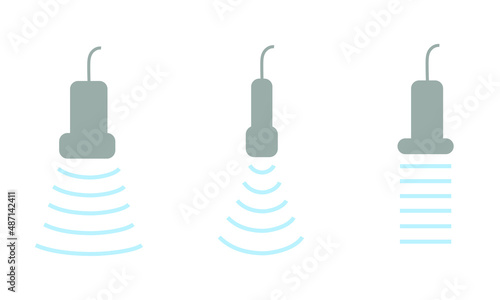
Three Ultrasound Transducer Including Curvilinear Cardiac and Linear Probe
Infographic of Ultrasound Probe Including Curvilinear and Transvaginal Transducer For Obstetrics and Gynecology. Medical Equipment
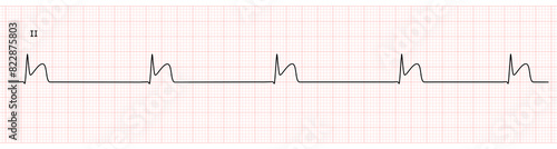
EKG Monitor Showing Sinus Rhythm with PVC
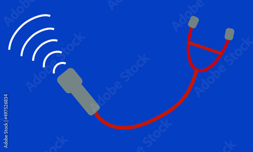
Ultrasound Probe with Stethoscope for Use as Point of care
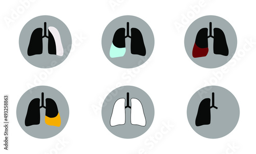
icon of lung and pleural pathology
EKG Monitor Showing Sinus Rhythm with Trigeminy PVC
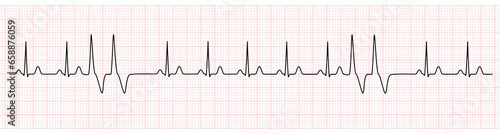
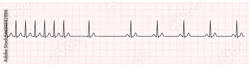
EKG Monitor Showing Atrial Fibrillation With Rapid Ventricular Response
pictogram of curvilinear or abdominal probe ultrasound. medical equipment concept
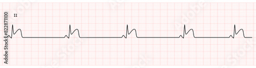
EKG Monitor Showing Sinus Rhythm with PAC
EKG Monitor Showing accelerated junctional rhythm
EKG Showing PR prolong which is Sign of First degree AV Block
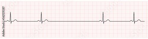
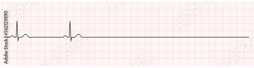
EKG Showing Sinus Bradycardia with pause of Patient
EKG Monitor in lead II Showing Sinus Bradycardia with STEMI at Inferior Wall
EKG Monitor in lead II Showing junctional Bradycardia with STEMI at Inferior Wall
point of care in cardiac ultrasound
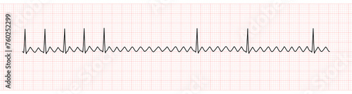
EKG Monitor Showing Atrial Flutter
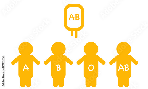
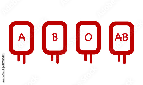
AB Blood Group of Plasma and Platelets. The Universal Donor on Which Background
EKG monitoring Show Ventricular Fibrillation
EKG Monitor Showing Sinus Rhythm with Short Run VT
EKG Monitor Showing Regularized Atrial Fibrillation or AF with Block
EKG Monitor Showing Ventricular tachycardia or VT
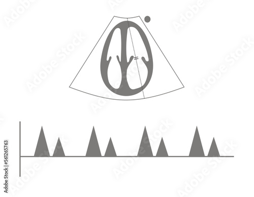
Infographic of Echocardiography of Left Ventricular Outflow Tract Velocity Time Integral or LVOT VTI From Apical Five Chamber View
EKG Showing Sinus Bradycardia with Arrest of Patient
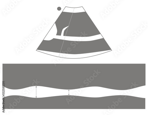
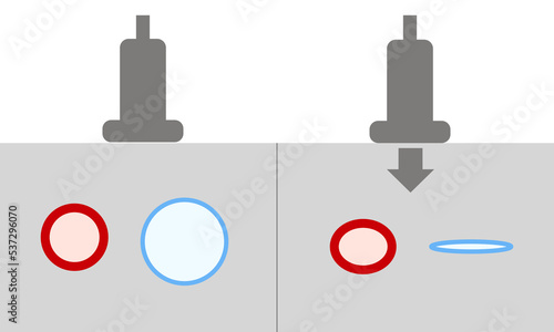
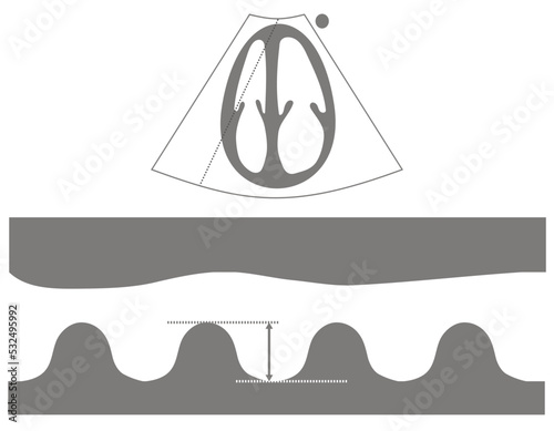
Diagram of Normal Compression Ultrasound For Diagnosis of Deep Vein Thrombosis
Ultrasound Probe with Stethoscope for Use as Point of care
Point of Care Ultrasound. New Tool for Use Instead of Stethoscope
Set of Icon of Extended FAST Examination positive
Pack Red Blood Cell of 4 ABO Blood Group on White Background
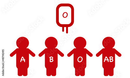
Pack Red Cell Group O. The Universal Blood Donor on White Background
multi shade of pink mountains and blue sky. pastel background concept
pictogram of position for emergency intubation in children. healthcare and medical concept
sniffing position for adult patient before emergency intubation
EKG Monitor in lead II Showing Complete heart block with STEMI at Inferior Wall
EKG Monitor Showing Wide QRS Complex From Hyperkalemia
EKG Monitor Showing Atrial Flutter with 2:1 AV Block After Adenosine Intravenous
EKG Monitor in lead II Showing Sinus Rhythm with STEMI at Inferior Wall
EKG Monitor Showing Atrial Flutter with 2:1 AV Block
EKG Monitor Showing Sinus Rhythm with U wave represented Hypokalemia
EKG Monitor Showing Sinus Rhythm with Tall T Wave Represented Hyperkalemia
EKG Monitor Showing Sinus Rhythm with U wave represented Hypokalemia
EKG Monitor Showing Sinus Rhythm with Couplets PVC
EKG Monitor Showing Sinus Rhythm with Bigeminy PVC
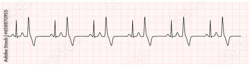
EKG Monitor Showing Atrial Fibrillation With Slow Ventricular Response
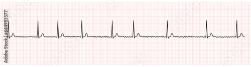
EKG Monitor Showing Atrial Fibrillation With Normal Ventricular Response
EKG Monitor Showing Atrial Fibrillation With Rapid Ventricular Response and Slower Rate After Adenosine Intravenous
Golden Circle Pattern on Red Background
Golden Circle Pattern on Red Background
Stripe of Pink Red and White Color Background
EKG Monitor Showing Supraventricular Tachycardia Change to Sinus Rhythm After Adenosine Injection
EKG Monitor Showing junctional escape rhythm
EKG Showing second degree av block mobitz II
Infographic of Echocardiogram of Apical Five Chamber And Measurement of LVOT VTI
EKG Showing second degree av block mobitz I
Infographic of Echocardiography of Left Ventricular Outflow Tract Velocity Time Integral or LVOT VTI From Apical Five Chamber View
Infographic of Ultrasound Transducer Including Linear Probe and Hockey Stick Probe for Musculoskeletal System Scan
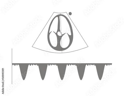
Infographic of Echocardiography of Mitral Inflow Velocity
Infographic of Intercostal Drainage System on White Background
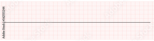
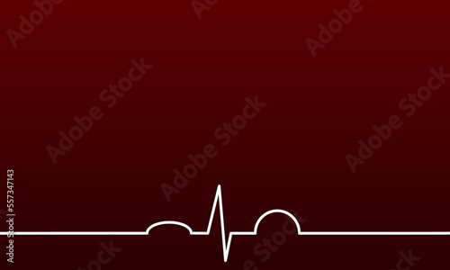
Red Background with normal EKG
Multicultural Face of People Talking Together
Ambulance Running on Road
Diagram of Abnormal Non Compression Ultrasound For Diagnosis of Deep Vein Thrombosis
Close up Different Shade of Hands Color Represent Diversity of People
Mixed Race of Children Sitting Together
Diagram of Inferior Vena Cava Ultrasound
Diagram of Measurement of Tricuspid Annulus Plane Systolic Excursion or TAPSE From Echocardiogram
Point of Care Ultrasound with Icon Showed Abnormal Findings
Diagram of Parasternal Long Axis View of Echocardiography. Healthcare Concept
Diagram of Subxiphoid View of Echocardiography. Healthcare Concept
Diagram of right Ventricle enlargement of Apical 4-Chamber View of Echocardiography. Healthcare Concept
Point of Care Ultrasound with Icon Showed Abnormal Findings
Diagram of Parasternal Short Axis View at Mitral Valve of Echocardiography. Healthcare Concept
Diagram of right Ventricle D Shape of Parasternal Short Axis View at Papillary Muscle of Echocardiography. Healthcare Concept
Diagram of Parasternal Short Axis View at Papillary Muscle of Echocardiography. Healthcare Concept
E-FAST with Icon Showing Abnormal Pathology
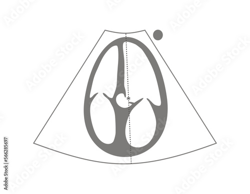
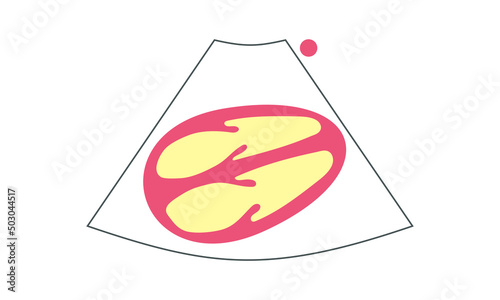
Diagram of Apical 4-Chamber View of Echocardiography. Healthcare Concept
Set of Icon of Normal Extended FAST Examination
Small Green Plant in Pot
Icon of Sonographic Finding of Gallbladder and Gallstone Pathology