Login
Sign Up
Member Area
Login
Sign Up
English
Afrikaans formal
Afrikaans informal
Arabic
Azerbaijan
Brazilian
Bulgarian
Catalan
Chinese simplified
Chinese traditional
Croatian
Czech
Danish
Dutch
English
Estonian
Finnish
French
Georgian
German
Greek
Hebrew
Hungarian
Icelandic
Indonesian
Italian
Japanese
Latvian
Lithuanian
Malaysian
Norwegian
Persian
Polish
Portuguese
Romanian
Russian
Serbian
Slovakian
Slovenian
Spanish
Swedish
Thai
Turkish
Ukrainian
Vietnamese
Toggle navigation
VideotonStock
Your Shopping Cart is empty.
Files
Stock
Photo
Video
Special offer
Collections
Packages
Lightboxes
Licenses
Royalty free
Rights managed
Creative
Editorial
Items
Most downloaded
Featured
Most popular
New
Free
Categories
Browse categories
Site info
Site info
About
Support
Privacy Policy
Privacy Policy
FAQ
Contact Us
Contact Us
Terms and Conditions
Buyer Agreement
Seller Agreement
Customers
Users
Credits
Photographers
Boby Richards
0
Boby Richards
0
Videoton1
286
Search
Keywords:
Creator ID:
Type:
Photo
Illustration
Vector
Video
Template
3d
Categories:
Animals
Buildings and Architecture
Business
Drinks
The Environment
States of Mind
Food
Graphic Resources
Hobbies and Leisure
Industry
Landscapes
Lifestyle
People
Plants and Flowers
Culture and Religion
Science
Social Issues
Sports
Technology
Transport
Travel
News
Entertainment
Sport News
Languages:
Brazilan Portuguese - Brazil
English - Canada
French - Canada
Spanish - Mexico
English - United States
English - Belgium
French - Belgium
Dutch - Belgium
Czech - Czech Republic
English - Cyprus
Danish - Danmark
German - Germany
English - Estonia
Spanish - Spain
French - France
French - Marocco
English - Greece
English - Ireland
Italian - Italia
English - Latvia
English - Lithuania
German - Luxembourg
English - Luxembourg
French - Luxembourg
English - Hungary
English - Malta
Dutch - Nederlands
Bokmal Norwegian - Norway
German - Austria
Polish - Poland
Portuguese - Portugal
English - Romania
German - Switzerland
English - Slovenia
Slovakian - Slovakia
French - Switzerland
English - Finland
Swedish- Sweden
Italian - Switzerland
English - United Kingdom
English - Bulgaria
English - Australia
Japanese - Japan
Korean - South Korea
English - New Zealand
Russian - Russia
English - Ukrain
English - Thailand
Orientation:
All
Horizontal
Vertical
square
Color:
Model property release:
All
Yes
No
Duration Video:
All
Up to 10 seconds
Up to 20 seconds
Longer than 30 seconds
Results
(0)
Site
Getty/iStock
Bigstockphoto
Pixabay
Adobe Stock
Relevance
New
Most popular
Most downloaded
Undiscovered
12
24
36
48
96
Auto paging
Menu
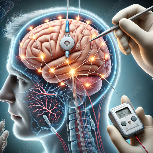
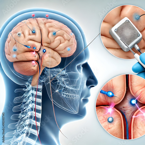
Brain anatomy: illustrating deep brain stimulation (DBS) therapy for a Parkinson’s patient. surgeon implanting electrodes into patient's brain, targeting specific areas like subthalamic nucleus.
Brain anatomy: illustrating deep brain stimulation (DBS) therapy for a Parkinson’s patient. surgeon implanting electrodes into patient's brain, targeting specific areas like subthalamic nucleus.
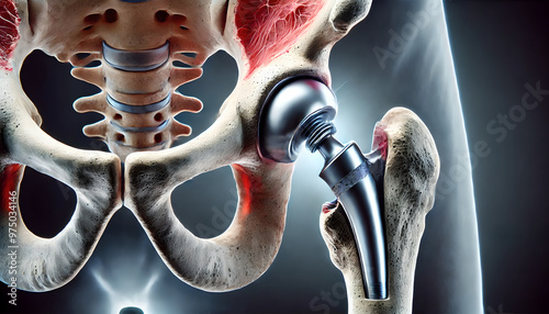
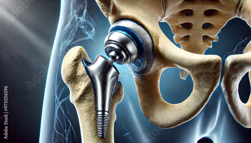
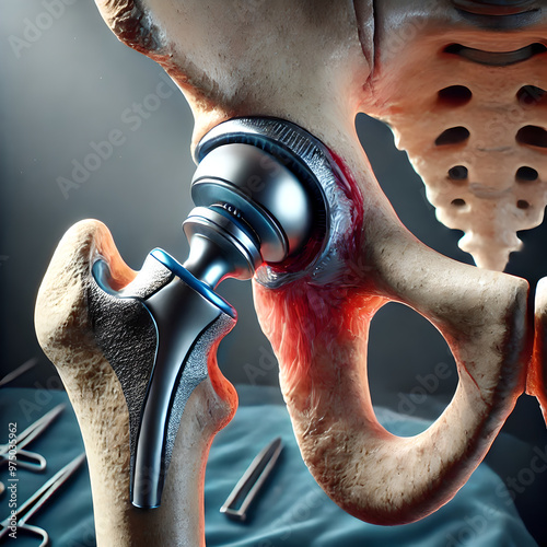
3D Bone: Hip replacement surgery, surgeon replacing damaged hip joint with prosthetic implant. focus on metal, plastic components of prosthetic, artificial ball and socket in place of natural joint.
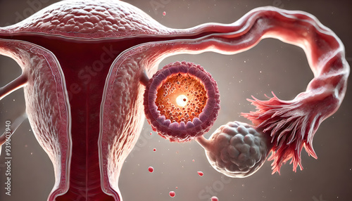
cross-sectional female ovary during ovulation phase menstrual cycle. follicle releasing an egg (oocyte) into the fallopian tube. structure of the ovary, egg towards tube.
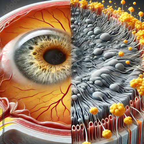
3D Age-related macular degeneration (AMD) in the human eye, focusing on the central portion of the retina where the degeneration occurs. accumulation of drusen (yellow deposits) under the retina.
Ear anatomy and intricate details of the inner ear, Ear drum, Ossicles, Auditory nerve - g
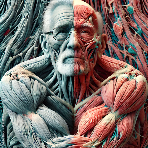
3D showing gradual degeneration of muscle tissue in elderly individuals, focusing reduction in muscle mass (sarcopenia), loss of muscle strength. muscle fibers, increase in connective tissue.
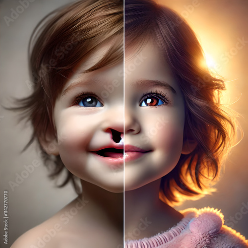
Comparison image showing a child before and after cleft lip and palate surgery. improvement in facial structure and overall appearance, highlighting the success of the corrective procedure.
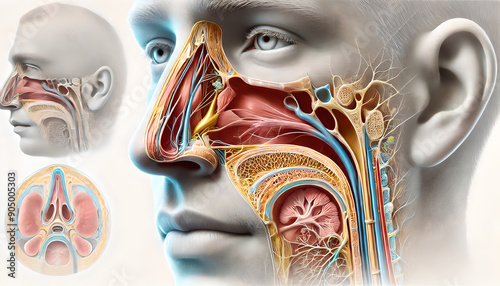
Anatomy of the Nose: Nasal anatomy, Health and Function in Respiratory and Olfactory Systems-d
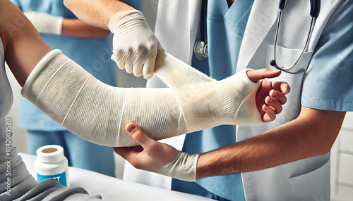
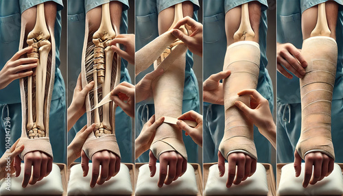
Healthcare professional applying cast to patient’s broken arm. show step-by-step process, including wrapping the soft padding around the arm, followed by application of the plaster or fiberglass cast.
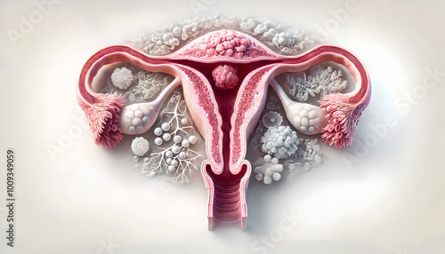
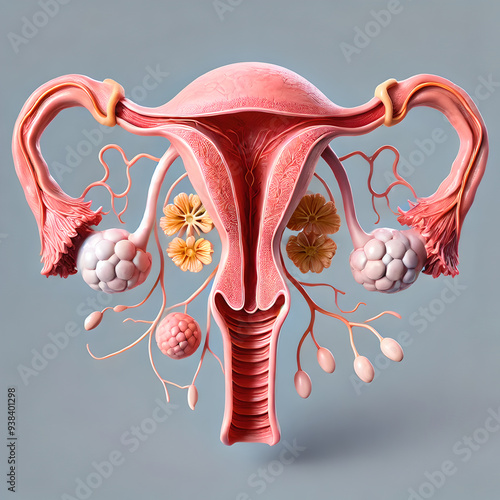
Uterine cancer. cross-section of uterus, layers endometrium, myometrium, cervix. Emphasize cancerous growths, tumors within endometrium, contrasting them with normal tissue.
Healthcare professional applying cast to patient’s broken arm. show step-by-step process, including wrapping the soft padding around the arm, followed by application of the plaster or fiberglass cast.
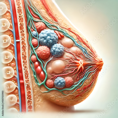
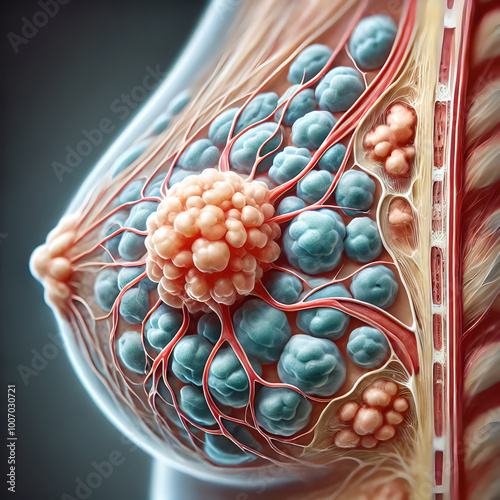
Image early stages of breast cancer, focusing on small, localized tumor developing within breast tissue. formation of abnormal cells in milk ducts (ductal carcinoma in situ), lobules.
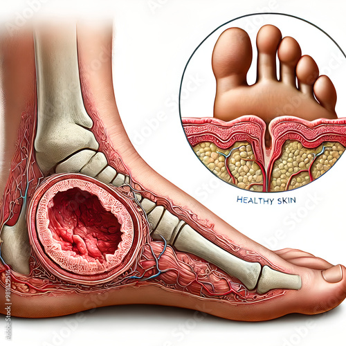
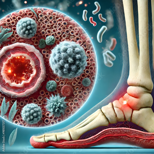
Foot anatomy: diabetic foot ulcer developing on the sole of the foot. ulcer’s location near the ball of the foot, with surrounding areas showing signs of infection or poor circulation.
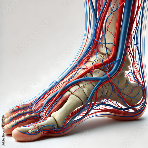
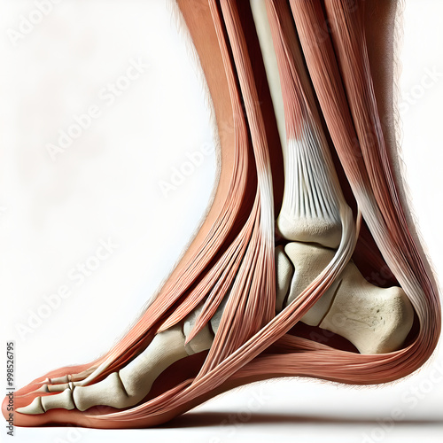
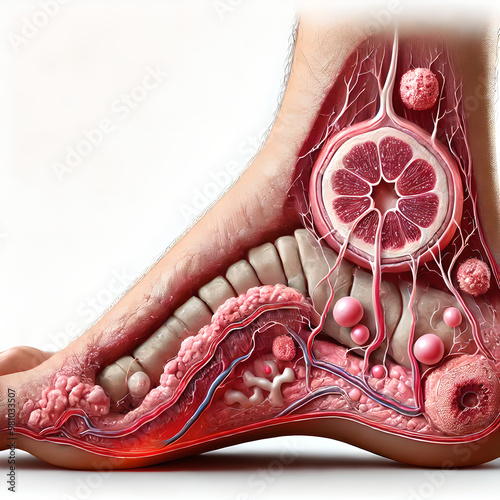
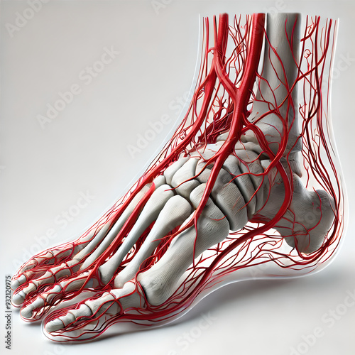
Detail human foot anatomy: emphasizing vascular and tendinous structures, medical, focusing on circulatory and musculoskeletal systems for educational purposes, realistic, blood vessels and tendons.
Uterine cancer. cross-section of uterus, layers endometrium, myometrium, cervix. Emphasize cancerous growths, tumors within endometrium, contrasting them with normal tissue.
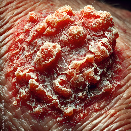
Squamous cell carcinoma (SCC) on a patient’s skin. show a rough, scaly patch or raised lesion, crusted over and sometimes ulcerated. Focus scaling, redness, and irregular thickness in affected area.
A realistic image patient with Parkinson’s disease experiencing tremors in their hands. focus on involuntary shaking, with a close-up of patient's hand to capture the subtle but persistent tremors.
3D model anatomy: Inflammation of the trachea (tracheitis), highlighting swelling and redness of mucous membrane lining trachea. mucus within airway and resulting narrowing of airway passage.
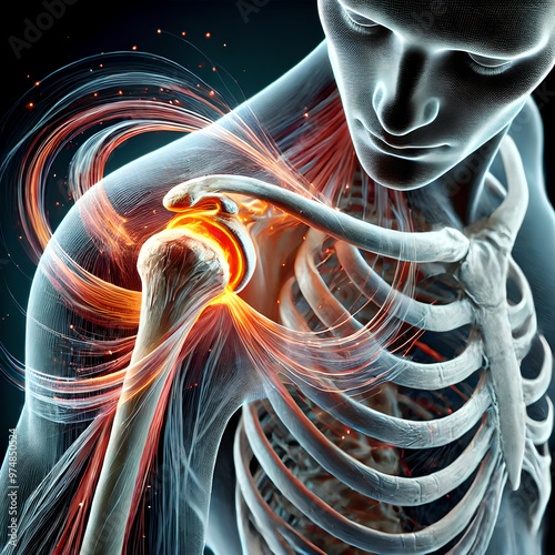
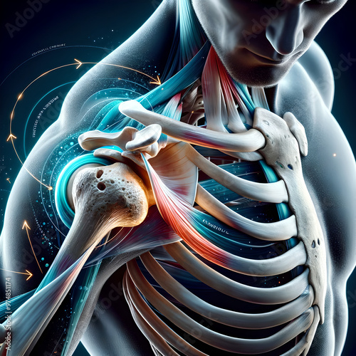
3D bone: human joint in motion, showing humerus, scapula, clavicle work together during shoulder rotation. movement of rotator cuff muscles, stabilize shoulder joint. synovial fluid in lubricating.
An AI Robot Creating Art by Drawing and Painting a Picture in the Air
cross-sectional female ovary during ovulation phase menstrual cycle. follicle releasing an egg (oocyte) into the fallopian tube. structure of the ovary, egg towards tube.
3D Bone: Hip replacement surgery, surgeon replacing damaged hip joint with prosthetic implant. focus on metal, plastic components of prosthetic, artificial ball and socket in place of natural joint.
3D showing gradual degeneration of muscle tissue in elderly individuals, focusing reduction in muscle mass (sarcopenia), loss of muscle strength. muscle fibers, increase in connective tissue.
3D showing how excess fat accumulates in various parts of the body in a person with obesity. buildup of fat around the abdomen, thighs, arms, and internal organs like the liver (visceral fat).
Brain anatomy: illustrating deep brain stimulation (DBS) therapy for a Parkinson’s patient. surgeon implanting electrodes into patient's brain, targeting specific areas like subthalamic nucleus.
A detailed image of a modern Intensive Care Unit (ICU) room equipped with advanced medical devices. hospital bed with multiple monitors displaying vital signs, ventilators, IV stands, infusion pumps.
The CT scan clearly shows kidney stones located in the renal pelvis. A contrast agent was used to make the stones highly visible against the surrounding tissue. The urinary tract is also visible.
3D Bone: Hip replacement surgery, surgeon replacing damaged hip joint with prosthetic implant. focus on metal, plastic components of prosthetic, artificial ball and socket in place of natural joint.
3D Bone: Hip replacement surgery, surgeon replacing damaged hip joint with prosthetic implant. focus on metal, plastic components of prosthetic, artificial ball and socket in place of natural joint.
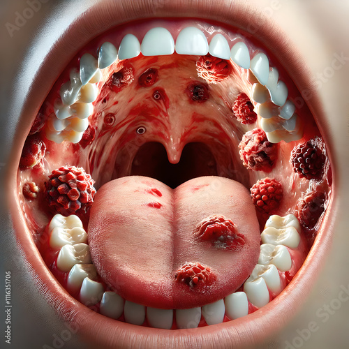
A close-up image of the inside of a child’s mouth showing painful ulcers caused by Hand, Foot, and Mouth Disease. highlight small, red sores on the tongue, gums, and inner cheeks.
3D Bone: Hip replacement surgery, surgeon replacing damaged hip joint with prosthetic implant. focus on metal, plastic components of prosthetic, artificial ball and socket in place of natural joint.
cross-sectional female ovary during ovulation phase menstrual cycle. follicle releasing an egg (oocyte) into the fallopian tube. structure of the ovary, egg towards tube.
3D bone: human joint in motion, showing humerus, scapula, clavicle work together during shoulder rotation. movement of rotator cuff muscles, stabilize shoulder joint. synovial fluid in lubricating.
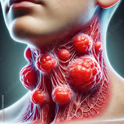
Close-up image swollen and inflamed lymph nodes due to infection or injury, case of lymphadenitis. focus on enlarged nodes in neck or armpits, showing redness, swelling in surrounding tissue.
Foot anatomy: diabetic foot ulcer developing on the sole of the foot. ulcer’s location near the ball of the foot, with surrounding areas showing signs of infection or poor circulation.
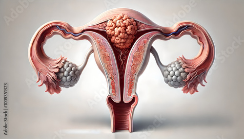
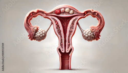
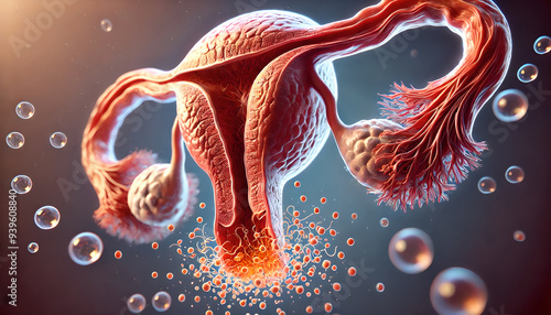
Detailed anatomy female reproductive system, highlighting uterus, ovaries, fallopian tubes, vagina, including endometrium, myometrium, perimetrium, along with the ovarian follicles at various stages.
Heart anatomy: 3D human heart illustrating coronary artery disease (CAD) with significant plaque build-up in the coronary arteries. focus highlight the narrowed areas of the arteries.
3D model illustration human Bone anatomy: high-resolution image of a knee joint for medical visualization, ball-and-socket joint to show its range of motion, medical education, diagram of a human hing
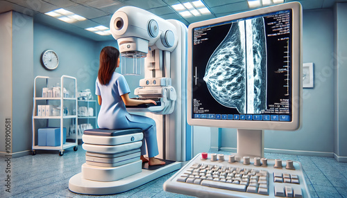
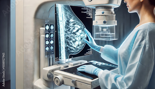
Mammogram being performed on patient to detect signs of breast cancer. breast tissue on mammogram screen, potential calcifications, masses, abnormal structures. Focus on the medical equipment.
Image early stages of breast cancer, focusing on small, localized tumor developing within breast tissue. formation of abnormal cells in milk ducts (ductal carcinoma in situ), lobules.
Foot anatomy: diabetic foot ulcer developing on the sole of the foot. ulcer’s location near the ball of the foot, with surrounding areas showing signs of infection or poor circulation.
3D bone: human joint in motion, showing humerus, scapula, clavicle work together during shoulder rotation. movement of rotator cuff muscles, stabilize shoulder joint. synovial fluid in lubricating.
3D Age-related macular degeneration (AMD) in the human eye, focusing on the central portion of the retina where the degeneration occurs. accumulation of drusen (yellow deposits) under the retina.
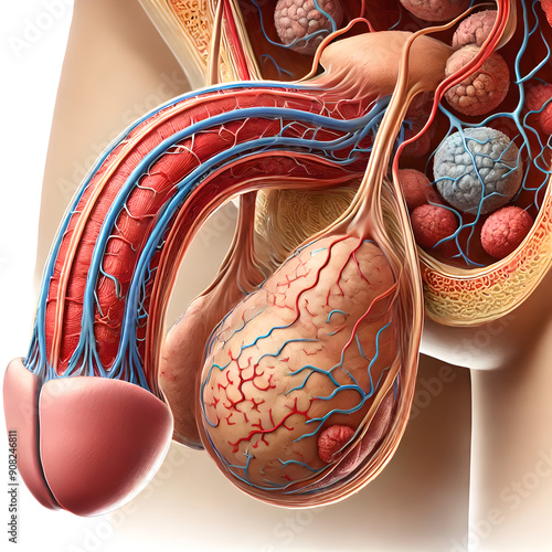
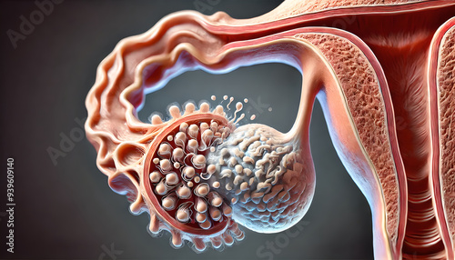
Detailed Overview of the Male Reproductive System: Focusing on the Anatomy and Function of the Testicle-c
Image comparison between high-calorie, balanced meal designed for weight loss with obesity. nutrient-rich foods, vegetables, lean protein, whole grains.
3D Age-related macular degeneration (AMD) in the human eye, focusing on the central portion of the retina where the degeneration occurs. accumulation of drusen (yellow deposits) under the retina.
cross-sectional female ovary during ovulation phase menstrual cycle. follicle releasing an egg (oocyte) into the fallopian tube. structure of the ovary, egg towards tube.
3D showing gradual degeneration of muscle tissue in elderly individuals, focusing reduction in muscle mass (sarcopenia), loss of muscle strength. muscle fibers, increase in connective tissue.
Uterine cancer. cross-section of uterus, layers endometrium, myometrium, cervix. Emphasize cancerous growths, tumors within endometrium, contrasting them with normal tissue.
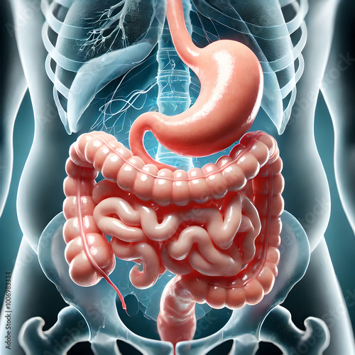
3D model surgical common bariatric surgery procedure, such as gastric bypass or sleeve gastrectomy. focus on the modification of the stomach to reduce its size, digestive system.
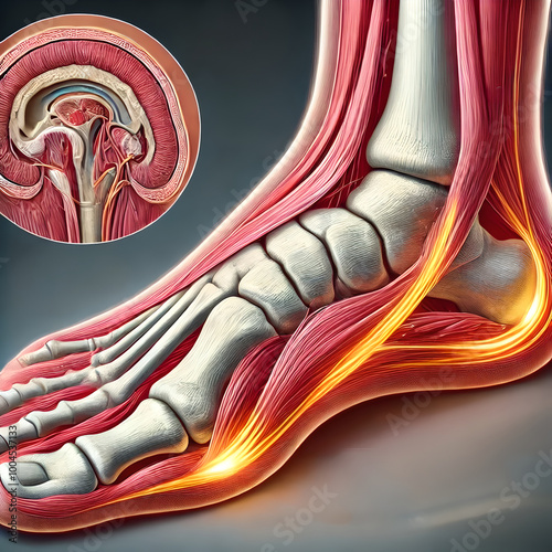
3D detailed plantar fascia tendon in the sole of foot, focusing on inflammation due to plantar fasciitis. thickening, tension in tendon, particularly near heel bone. one affected by plantar fasciitis.
3D detailed plantar fascia tendon in the sole of foot, focusing on inflammation due to plantar fasciitis. thickening, tension in tendon, particularly near heel bone. one affected by plantar fasciitis.
3D detailed plantar fascia tendon in the sole of foot, focusing on inflammation due to plantar fasciitis. thickening, tension in tendon, particularly near heel bone. one affected by plantar fasciitis.
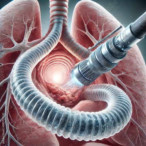
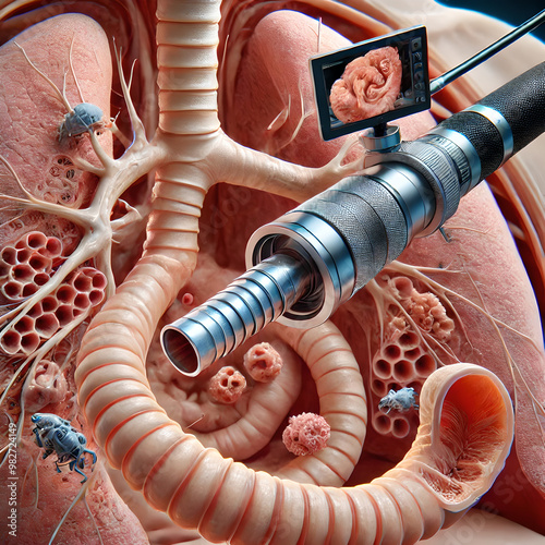
3D Model human trachea anatomy: process of bronchoscopy, where a flexible camera is inserted into the trachea to examine the airway. tracheal walls, mucus, any potential blockages or abnormalities.
3D Age-related macular degeneration (AMD) in the human eye, focusing on the central portion of the retina where the degeneration occurs. accumulation of drusen (yellow deposits) under the retina.
Human kidney with acute glomerulonephritis, showing inflammation ,swelling glomeruli. visibility inflamed areas within nephron, highlighting damage to filtering units kidney. inflamed, fibrotic tissue
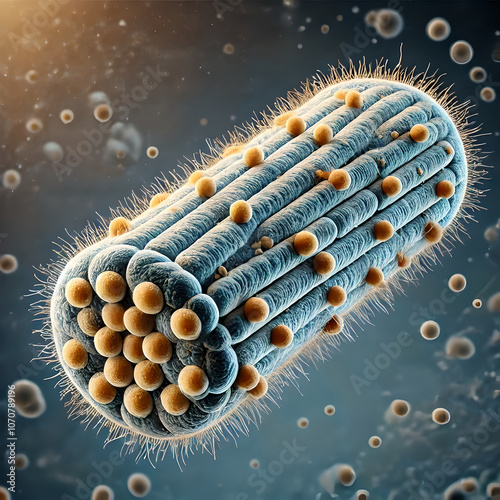
Microscopic image of Clostridium tetani bacteria, the pathogen responsible for tetanus infection. focus on rod-shaped bacteria with terminal spores, neurotoxin, causes muscle spasms and stiffness.
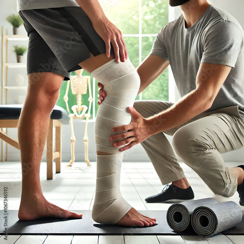
Patient undergoing physical therapy for leg strength and mobility recovery after having a cast removed. focus on exercises and stretches to regain range of motion in the knee and ankle.
Healthy skin versus skin affected by basal cell carcinoma (BCC). smooth, even texture of healthy skin alongside raised, pearly lesion with visible blood vessels or central depression.
Healthcare professional applying cast to patient’s broken arm. show step-by-step process, including wrapping the soft padding around the arm, followed by application of the plaster or fiberglass cast.
Lymph node biopsy procedure, where a small portion or the entire lymph node is removed for diagnostic testing. focus on the incision site and the tools used to extract the lymph node.
Close-up image swollen and inflamed lymph nodes due to infection or injury, case of lymphadenitis. focus on enlarged nodes in neck or armpits, showing redness, swelling in surrounding tissue.
Close-up image swollen and inflamed lymph nodes due to infection or injury, case of lymphadenitis. focus on enlarged nodes in neck or armpits, showing redness, swelling in surrounding tissue.
Close-up image swollen and inflamed lymph nodes due to infection or injury, case of lymphadenitis. focus on enlarged nodes in neck or armpits, showing redness, swelling in surrounding tissue.
Close-up image swollen and inflamed lymph nodes due to infection or injury, case of lymphadenitis. focus on enlarged nodes in neck or armpits, showing redness, swelling in surrounding tissue.
Anatomical diagram structure and location of lymph nodes throughout human body. major clusters of lymph nodes in areas such as neck (cervical), armpits (axillary), groin (inguinal), abdomen.
Breeding grounds of Aedes mosquitoes. Include stagnant water sources like puddles, flowerpots, and water containers, with adult mosquitoes laying eggs in these environments.
Breeding grounds of Aedes mosquitoes. Include stagnant water sources like puddles, flowerpots, and water containers, with adult mosquitoes laying eggs in these environments.
Uterine cancer. cross-section of uterus, layers endometrium, myometrium, cervix. Emphasize cancerous growths, tumors within endometrium, contrasting them with normal tissue.
Mammogram being performed on patient to detect signs of breast cancer. breast tissue on mammogram screen, potential calcifications, masses, abnormal structures. Focus on the medical equipment.
Mammogram being performed on patient to detect signs of breast cancer. breast tissue on mammogram screen, potential calcifications, masses, abnormal structures. Focus on the medical equipment.
Image early stages of breast cancer, focusing on small, localized tumor developing within breast tissue. formation of abnormal cells in milk ducts (ductal carcinoma in situ), lobules.
Image early stages of breast cancer, focusing on small, localized tumor developing within breast tissue. formation of abnormal cells in milk ducts (ductal carcinoma in situ), lobules.
Image comparison between high-calorie, balanced meal designed for weight loss with obesity. nutrient-rich foods, vegetables, lean protein, whole grains.
3D model surgical common bariatric surgery procedure, such as gastric bypass or sleeve gastrectomy. focus on the modification of the stomach to reduce its size, digestive system.
3D model surgical common bariatric surgery procedure, such as gastric bypass or sleeve gastrectomy. focus on the modification of the stomach to reduce its size, digestive system.
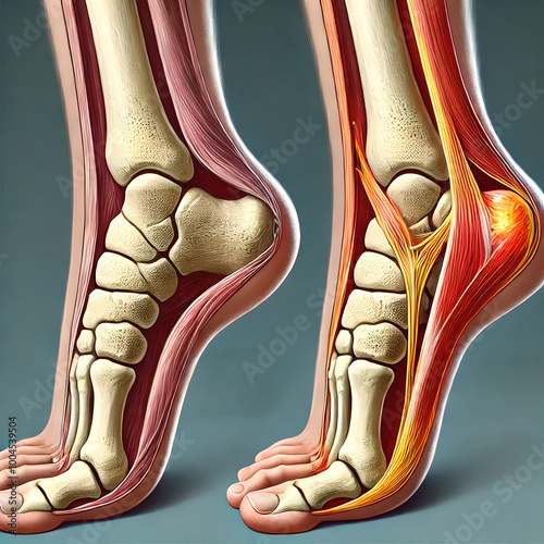
A detailed anatomical diagram of the Achilles tendon, showing its connection between the calf muscles (gastrocnemius and soleus), heel bone (calcaneus). thick, fibrous nature of tendon.
A detailed anatomical diagram of the Achilles tendon, showing its connection between the calf muscles (gastrocnemius and soleus), heel bone (calcaneus). thick, fibrous nature of tendon.
Capillaries surrounding the alveoli in lungs, illustrating process of gas exchange. oxygen moving from alveoli into the capillaries and carbon dioxide moving from the blood into the alveoli.
Close-up the effects of diabetes on the major arteries, with a focus on the aorta. thickening and hardening of the arterial walls (atherosclerosis), caused by prolonged high blood sugar levels.
3D Model human trachea anatomy: process of bronchoscopy, where a flexible camera is inserted into the trachea to examine the airway. tracheal walls, mucus, any potential blockages or abnormalities.
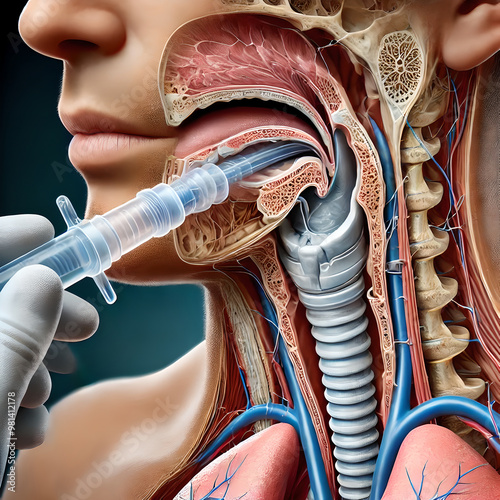
3D Model tracheal anatomy: tracheal intubation, where a breathing tube is inserted into the trachea to maintain an open airway. tube passing through the mouth and larynx into the trachea.
3D Model tracheal anatomy: tracheal intubation, where a breathing tube is inserted into the trachea to maintain an open airway. tube passing through the mouth and larynx into the trachea.
Foot anatomy: diabetic foot ulcer developing on the sole of the foot. ulcer’s location near the ball of the foot, with surrounding areas showing signs of infection or poor circulation.
Foot anatomy: diabetic foot ulcer developing on the sole of the foot. ulcer’s location near the ball of the foot, with surrounding areas showing signs of infection or poor circulation.
Foot anatomy: diabetic foot ulcer developing on the sole of the foot. ulcer’s location near the ball of the foot, with surrounding areas showing signs of infection or poor circulation.
3D Bone X-ray: showing structure of human knee joint. highlight bones, cartilage, joint space, showing signs of potential damage or injury. Focus reveal fractures, arthritis, or joint misalignments.
3D Bone: Hip replacement surgery, surgeon replacing damaged hip joint with prosthetic implant. focus on metal, plastic components of prosthetic, artificial ball and socket in place of natural joint.
3D bone: human joint in motion, showing humerus, scapula, clavicle work together during shoulder rotation. movement of rotator cuff muscles, stabilize shoulder joint. synovial fluid in lubricating.
3D showing gradual degeneration of muscle tissue in elderly individuals, focusing reduction in muscle mass (sarcopenia), loss of muscle strength. muscle fibers, increase in connective tissue.
3D Age-related macular degeneration (AMD) in the human eye, focusing on the central portion of the retina where the degeneration occurs. accumulation of drusen (yellow deposits) under the retina.
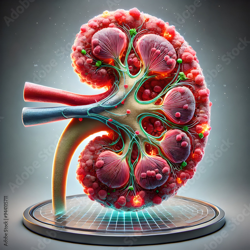
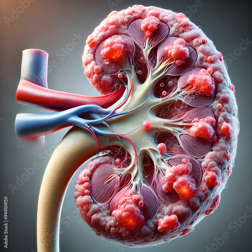
Human kidney with acute glomerulonephritis, showing inflammation ,swelling glomeruli. visibility inflamed areas within nephron, highlighting damage to filtering units kidney. inflamed, fibrotic tissue
Human kidney with acute glomerulonephritis, showing inflammation ,swelling glomeruli. visibility inflamed areas within nephron, highlighting damage to filtering units kidney. inflamed, fibrotic tissue
Human kidney with acute glomerulonephritis, showing inflammation ,swelling glomeruli. visibility inflamed areas within nephron, highlighting damage to filtering units kidney. inflamed, fibrotic tissue
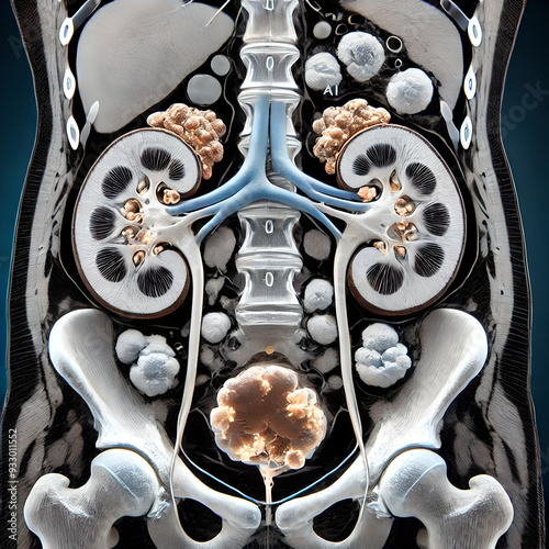
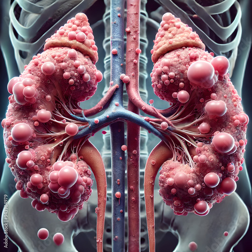
Human kidneys affected by polycystic kidney disease (PKD), showing numerous cysts varying sizes spread throughout tissue. swollen, irregularly shaped, cysts visibly displacing normal kidney structures
cross-sectional female ovary during ovulation phase menstrual cycle. follicle releasing an egg (oocyte) into the fallopian tube. structure of the ovary, egg towards tube.
cross-sectional female ovary during ovulation phase menstrual cycle. follicle releasing an egg (oocyte) into the fallopian tube. structure of the ovary, egg towards tube.
Detail human foot anatomy: emphasizing vascular and tendinous structures, medical, focusing on circulatory and musculoskeletal systems for educational purposes, realistic, blood vessels and tendons.
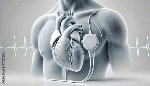
3D human heart with a pacemaker, clearly showing the device implanted.