Login
Sign Up
Member Area
Login
Sign Up
English
Afrikaans formal
Afrikaans informal
Arabic
Azerbaijan
Brazilian
Bulgarian
Catalan
Chinese simplified
Chinese traditional
Croatian
Czech
Danish
Dutch
English
Estonian
Finnish
French
Georgian
German
Greek
Hebrew
Hungarian
Icelandic
Indonesian
Italian
Japanese
Latvian
Lithuanian
Malaysian
Norwegian
Persian
Polish
Portuguese
Romanian
Russian
Serbian
Slovakian
Slovenian
Spanish
Swedish
Thai
Turkish
Ukrainian
Vietnamese
Toggle navigation
VideotonStock
Your Shopping Cart is empty.
Files
Stock
Photo
Video
Special offer
Collections
Packages
Lightboxes
Licenses
Royalty free
Rights managed
Creative
Editorial
Items
Most downloaded
Featured
Most popular
New
Free
Categories
Browse categories
Site info
Site info
About
Support
Privacy Policy
Privacy Policy
FAQ
Contact Us
Contact Us
Terms and Conditions
Buyer Agreement
Seller Agreement
Customers
Users
Credits
Photographers
Boby Richards
0
Boby Richards
0
Videoton1
286
Search
Keywords:
Creator ID:
Type:
Photo
Illustration
Vector
Video
Template
3d
Categories:
Animals
Buildings and Architecture
Business
Drinks
The Environment
States of Mind
Food
Graphic Resources
Hobbies and Leisure
Industry
Landscapes
Lifestyle
People
Plants and Flowers
Culture and Religion
Science
Social Issues
Sports
Technology
Transport
Travel
News
Entertainment
Sport News
Languages:
Brazilan Portuguese - Brazil
English - Canada
French - Canada
Spanish - Mexico
English - United States
English - Belgium
French - Belgium
Dutch - Belgium
Czech - Czech Republic
English - Cyprus
Danish - Danmark
German - Germany
English - Estonia
Spanish - Spain
French - France
French - Marocco
English - Greece
English - Ireland
Italian - Italia
English - Latvia
English - Lithuania
German - Luxembourg
English - Luxembourg
French - Luxembourg
English - Hungary
English - Malta
Dutch - Nederlands
Bokmal Norwegian - Norway
German - Austria
Polish - Poland
Portuguese - Portugal
English - Romania
German - Switzerland
English - Slovenia
Slovakian - Slovakia
French - Switzerland
English - Finland
Swedish- Sweden
Italian - Switzerland
English - United Kingdom
English - Bulgaria
English - Australia
Japanese - Japan
Korean - South Korea
English - New Zealand
Russian - Russia
English - Ukrain
English - Thailand
Orientation:
All
Horizontal
Vertical
square
Color:
Model property release:
All
Yes
No
Duration Video:
All
Up to 10 seconds
Up to 20 seconds
Longer than 30 seconds
Results
(0)
Site
Getty/iStock
Bigstockphoto
Pixabay
Adobe Stock
Relevance
New
Most popular
Most downloaded
Undiscovered
12
24
36
48
96
Auto paging
Menu
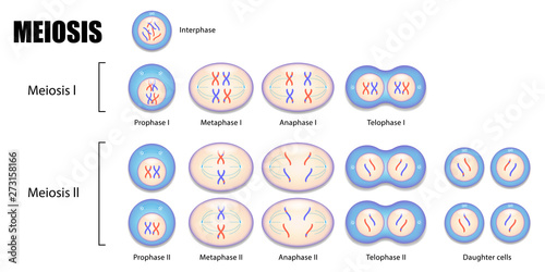
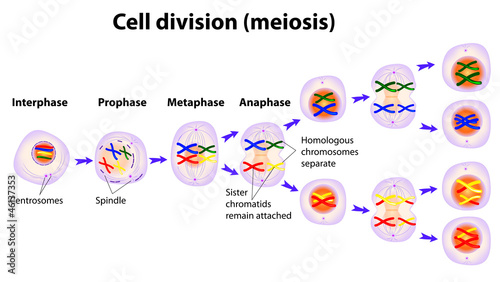
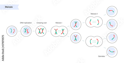
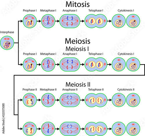
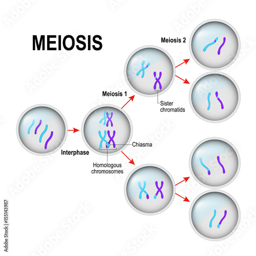
Diagram of Meiosis vector ESP10
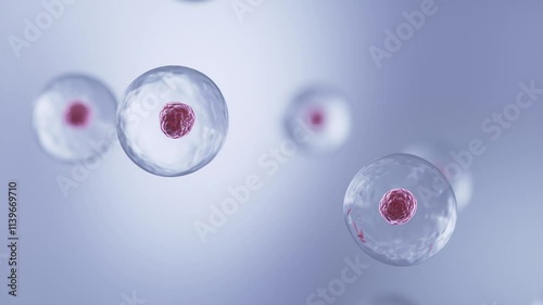
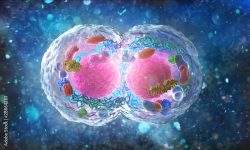
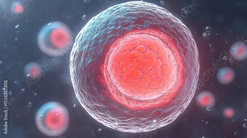
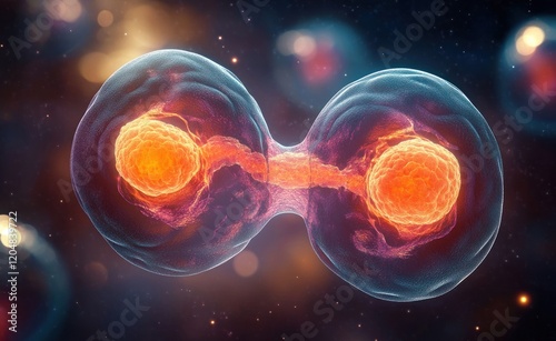
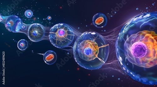
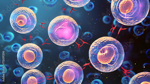
Human Cell division, cells under a microscope 3d animation.
Vector illustration of Meiosis phases. Cell division
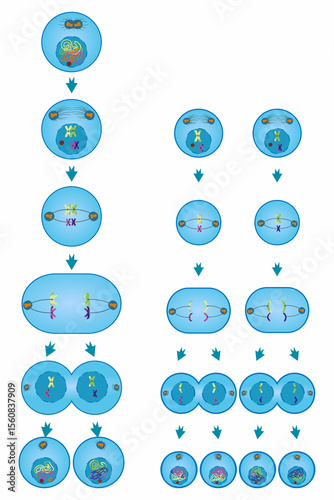
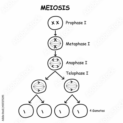
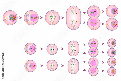
Diagram of Meiosis.Cell division is the process cells go through to divide.
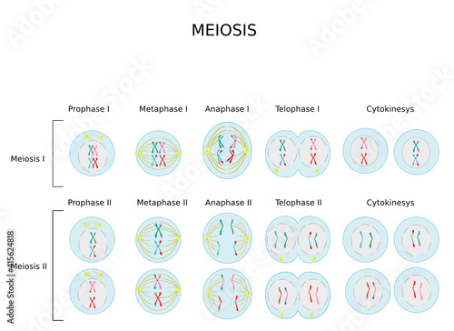
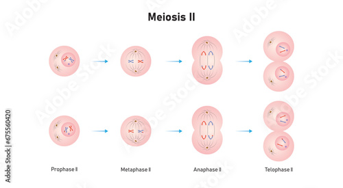
Meiosis. Meiotic division of an animal cell. Prophase, Metaphase, Anaphase, and Telophase.
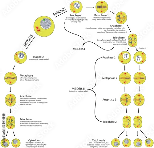
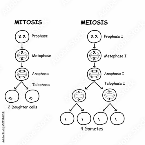
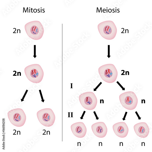
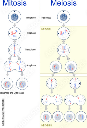
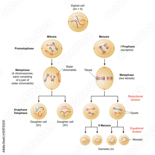
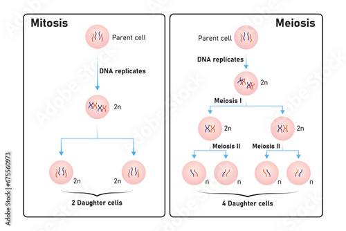
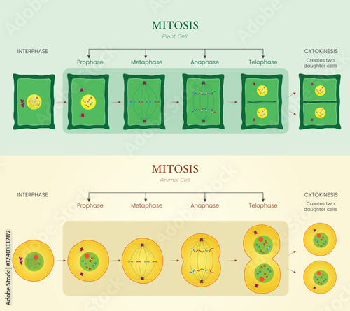
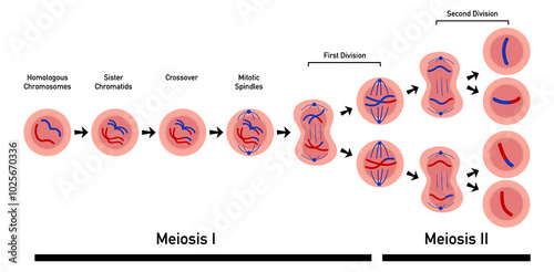
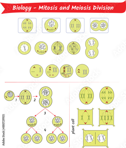
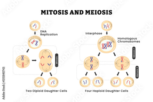
Mitosis and meiosis comparison of phases scheme with explanation
Microscopic View of Two Cells Connecting and Exchanging Genetic Material. Seamless Looping 4K Animation for Science, Medical, and Educational Videos. Cell Biology, Genetics, and Microbiology Concepts.
Animation of phases of meiosis with crossing-over
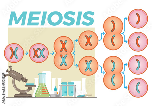
Cell division meiosis stages infographic diagram for biology science education cartoon vector drawing illustration sexually reproducing organism gametes chromosome reductional division of germ cells
Vector diagram of the meiosis phases
A surreal view of meiosis
Meiotic phases: Prophase, Metaphase, Anaphase, and Telophase.Process cell division in sexually reproducing organisms.Educational infographic
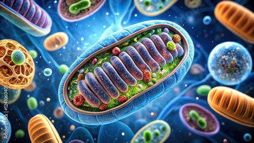
Mitochondria cell structure, meiosis, cellular metabolism, meiosis
Stages of meiosis vector illustration. Labeled cell division process scheme
mitosis and meiosis diagram. cell division
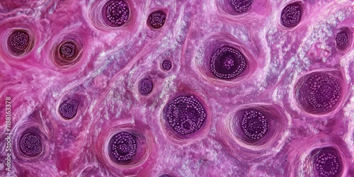
Fetal ovary micrograph showcasing multiple oocytes in the pachytene stage. The thick strands of paired homologous chromosomes are prominently visible in this detailed view of oocyte meiosis.
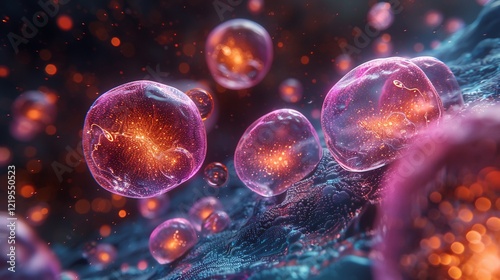
Colorful representation of a cell structure.
Meiosis of germ cells
Glowing neon Dna Structure icon isolated on black background. HD Video motion graphic animation
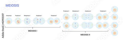
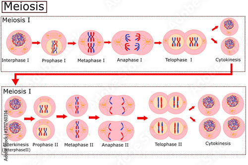
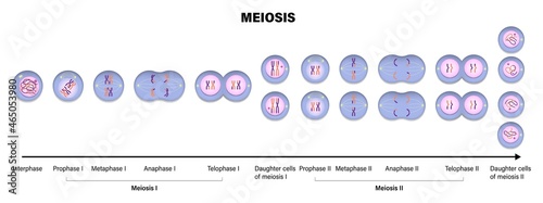
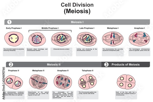
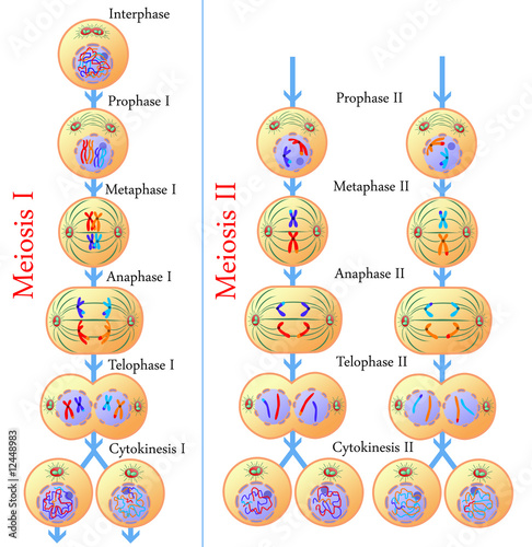
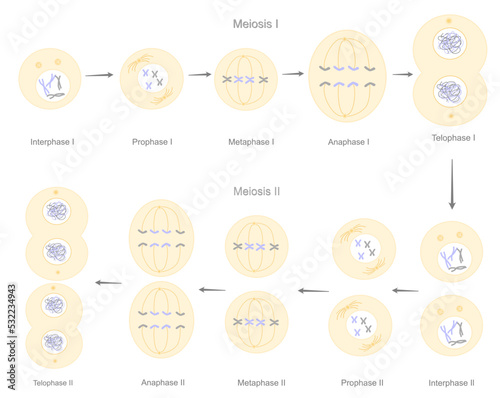
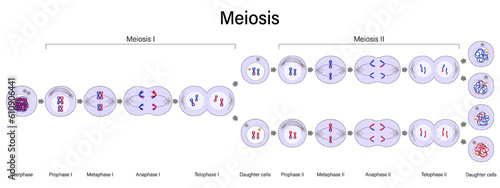
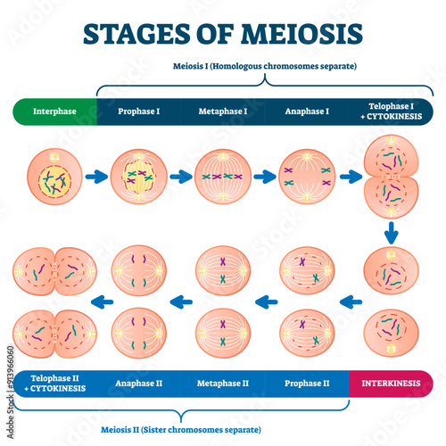
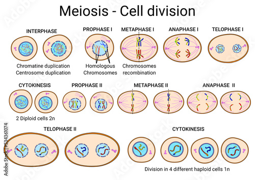
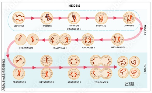
The Meiosis stages of reproductive cell division that contain the meiosis l and ll process : Interphase, Prophase, Metaphase, Anaphase and Telophase
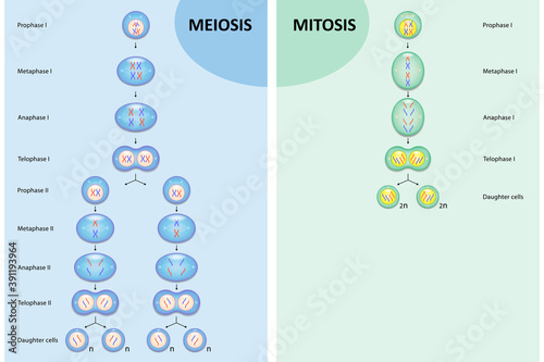
Diagram comparison of Meiosis and Mitosis, Process cell division
3D Animation of Cell division under a microscope. Cloning Cells. Cell mitosis concept.
Mitosis vs Meiosis Design Vector Illustration
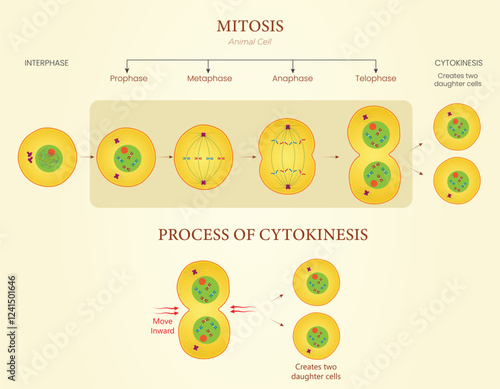
Diagram of Mitosis and Cytokinesis Illustration Showing Cell Division in Animal Cells
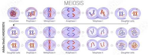
Diagram of Meiosis vector. Meiosis I and Meiosis II. Crossing over. Prophase, Metaphase, Anaphase, and Telophase. Cell division for education use.
Cell cycle and stages in division and meiosis of eukaryotic cell, 3D illustration
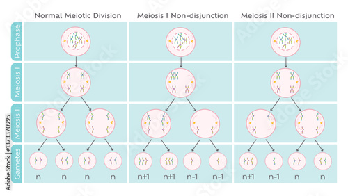
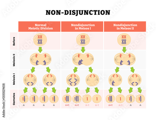
Nondisjunction in meiotic cell division educational diagram
Mitosis versus meiosis
Cells with Colorful Particles. Microscopic View of Biological Processes, Medical Visualization, and Scientific Research. Seamless Looping 4K Animation
Cell division. mitosis and meiosis
Meiosis cell division
Meiosis stage 2 (second polar body, and a single haploid cell, which enlarges to become an ovum)
Biology concept. Cell division under the microscope. 3d illustration
Schematic illustration with meiosis cell division process and various lab objects in the background
Cells divide by two mechanisms called mitosis and meiosis
Centrosome during anaphase 1 meiosis, divide into centrioles, spindle fibers originating from the centrosome participate in chromatid separation.
The Amazing Journey of Cell Division
Mitosis and Meiosis cell division
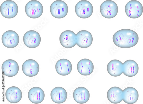
Stages of meiosis
cell biology
Meiosis I, First Division of Meiosis Science Design. Vector Illustration.
Microscopic Yeast Cell Division: Budding Process in High Resolution
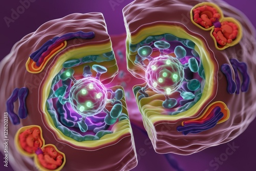
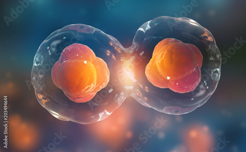
Cross-section view of a cell undergoing cytokinesis, showing the division of cytoplasm and organelles.
Diagram of Mitosis and Cytokinesis Illustration Showing Cell Division in Animal Cells
Diagram of Meiosis vector. Meiosis I and Meiosis II. Crossing over. Prophase, Metaphase, Anaphase, and Telophase. Cell division for education use.
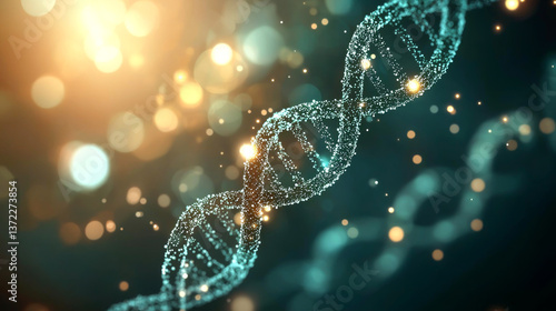
DNA Replication: Unraveling the Double Helix - Microscopic View
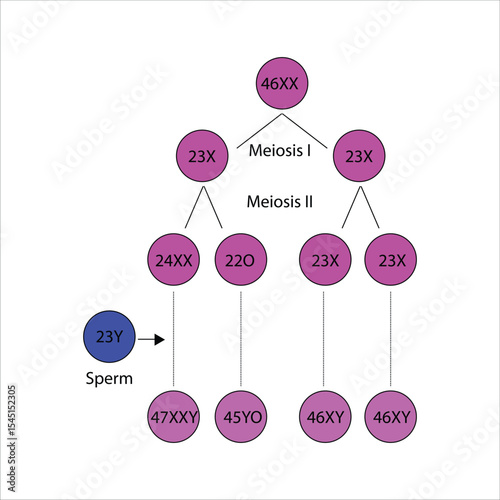
Chromosomal abnormalities in meiosis - Klinefelter's syndrome
Mitosis and Meiosis Scientific Design. Vector Illustration.
Cellular Development: Early Embryo Stages
meiosis. Cell division. In meiosis, the homologous chromosomes exchange genetic information
diagram of the stages of meiosis in animal cells
A high-tech molecular biology lab with digital screens displaying the structures of proteins and nucleic acids in 3D. 423
Cell division process showing cleavage and blastocyst formation
Stages of meiosis illustration, transparent background. Labeled cell division process explanation scheme from genetic aspect. Interphase and interkinesis diagram with phases structural changes.
Meiosis
Microscopic View of Cellular Fusion and Division in Abstract Art
Image of In Vitro Fertilization (IVF) Procedure in High-Tech Laboratory Setting
Cellular Development and Growth
Cell examination via microscope; depiction of cell division; cellular therapy shown; 3D image rendered on dark background
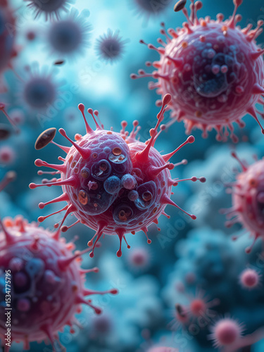
Magnification view of cell division and scattered Clostridium botulinum under microscope 3d rendering
Metaphase, second stage of meiosis and mitosis science design. vector Illustration.
Microscopic view of cellular division with glowing organelles.
Meiosis
Illustration of Mitosis Diagram Showing Cell Division in Plant and Animal Cells
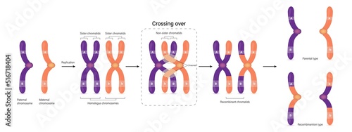
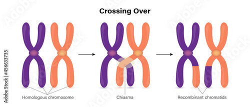
Diagram of Crossing over. Parental type and Recombination type. Genetic variation. Vector used for scientific and medical education.
3D Animation of Cell division under a microscope. Dividing and Multiplying Cells. Cell mitosis concept. Inside Human Body Animated.
Non Disjunction in meiosis diagram stages, labeled gametes, meiosis 1, 2, chromosomes or chromatids fail to separate during cell division, formation of genetic diseases. Vector illustration.
Easter bouquet
Meiosis 2 Phases Scientific Design. Vector Illustration.
Meiosis, cell division, homologous chromosomes, Meiosis I and Meiosis II, Crossing over, Prophase, Metaphase, Anaphase, and Telophase, Cell division for education use, Animation of phases
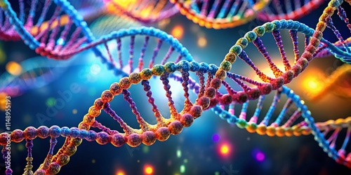
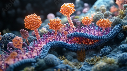
Abstract representation of DNA strands with glowing particles in the background.
diagram of the stages of meiosis in animal cells
diagram of meiosis cell division
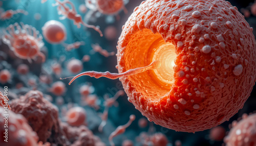
Microscopic view of a sperm penetrating an egg, surrounded by other cells. Detailed rendering showcases the fertilization process.
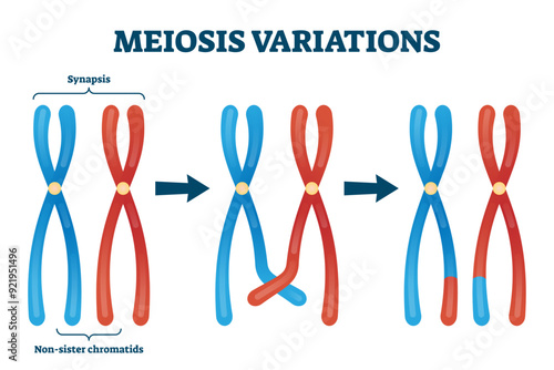
Meiosis variations illustration, transparent background. Educational genetic cell division. Scheme with synapsis and non sister chromatids. Diagram with meiosis stages and duplication.
Diakinesis is the last stage of meiosis prophase-1 (a five-stage process). Diakinesis stage is characterized by chiasmata terminalization.
Molecular marvel demonstrating mitosis meiosis compared high resolution detailing of chromosomal alignment under vibrant ambient holographic glow vibrant cinematic blending perfect accuracy designed
Meiosis. Cell division
Humans spermatogonis oogenesis scheme education vector illustration info graphic.
Meiosis cycle cell division School supplies Biology study
Mitosis and Meiosis Division in Cells
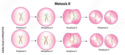
Meiosis II, Second Division of Meiosis Science Design. Vector Illustration.
Dramatic Portrayal of the Divine Dance of Life: The Formation of a Zygote
the stages of meiosis, from meiosis i to meiosis , cell, chromosomes, crossing over, homologous chromosomes, cell biology, meiosis, cell division, genetic variation, homologous chromosome, science, bi
Colorful illustration of homologous chromosomes, identical in shape and size, paired together with DNA double helix structure, demonstrating meiosis and genetic inheritance concept.
Crossing over of chromosome. Genetic recombination.
Mitosis and meiosis division, stages, plant cell.
Diagram showing the stages of meiosis with detailed annotations
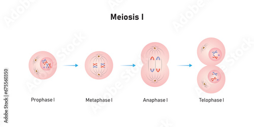
Meiosis 1 Phases Scientific Design. Vector Illustration.
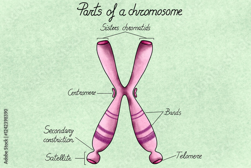
Anatomy of a Chromosome - Illustration
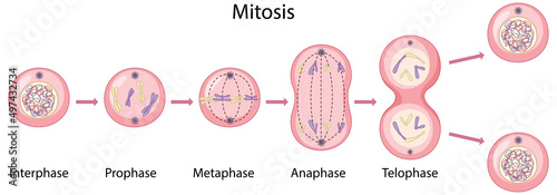
Mitosis and meiosis diagram. Cell division. Comparison diagram, healthcare poster vector illustration. Labeled Prophase, Metaphase, Anaphase, Interphase and Telophase, DNA replication.
Asexual reproduction fragmentation diagram
diagram of stages of meiosis
Meiosis of germ cells
Cell division in meiosis Telophase II observed under a microscope
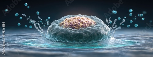
Cells under a microscope. Cell division. Cellular Therapy. 3d illustration on a dark background
Meiosis
Meiosis stage 1 (prophase, metaphase, anaphase and telophase stages in biology)
Cell division background
Process division of cell