Login
Sign Up
Member Area
Login
Sign Up
English
Afrikaans formal
Afrikaans informal
Arabic
Azerbaijan
Brazilian
Bulgarian
Catalan
Chinese simplified
Chinese traditional
Croatian
Czech
Danish
Dutch
English
Estonian
Finnish
French
Georgian
German
Greek
Hebrew
Hungarian
Icelandic
Indonesian
Italian
Japanese
Latvian
Lithuanian
Malaysian
Norwegian
Persian
Polish
Portuguese
Romanian
Russian
Serbian
Slovakian
Slovenian
Spanish
Swedish
Thai
Turkish
Ukrainian
Vietnamese
Toggle navigation
VideotonStock
Your Shopping Cart is empty.
Files
Stock
Photo
Video
Special offer
Collections
Packages
Lightboxes
Licenses
Royalty free
Rights managed
Creative
Editorial
Items
Most downloaded
Featured
Most popular
New
Free
Categories
Browse categories
Site info
Site info
About
Support
Privacy Policy
Privacy Policy
FAQ
Contact Us
Contact Us
Terms and Conditions
Buyer Agreement
Seller Agreement
Customers
Users
Credits
Photographers
Boby Richards
0
Boby Richards
0
Videoton1
286
Search
Keywords:
Creator ID:
Type:
Photo
Illustration
Vector
Video
Template
3d
Categories:
Animals
Buildings and Architecture
Business
Drinks
The Environment
States of Mind
Food
Graphic Resources
Hobbies and Leisure
Industry
Landscapes
Lifestyle
People
Plants and Flowers
Culture and Religion
Science
Social Issues
Sports
Technology
Transport
Travel
News
Entertainment
Sport News
Languages:
Brazilan Portuguese - Brazil
English - Canada
French - Canada
Spanish - Mexico
English - United States
English - Belgium
French - Belgium
Dutch - Belgium
Czech - Czech Republic
English - Cyprus
Danish - Danmark
German - Germany
English - Estonia
Spanish - Spain
French - France
French - Marocco
English - Greece
English - Ireland
Italian - Italia
English - Latvia
English - Lithuania
German - Luxembourg
English - Luxembourg
French - Luxembourg
English - Hungary
English - Malta
Dutch - Nederlands
Bokmal Norwegian - Norway
German - Austria
Polish - Poland
Portuguese - Portugal
English - Romania
German - Switzerland
English - Slovenia
Slovakian - Slovakia
French - Switzerland
English - Finland
Swedish- Sweden
Italian - Switzerland
English - United Kingdom
English - Bulgaria
English - Australia
Japanese - Japan
Korean - South Korea
English - New Zealand
Russian - Russia
English - Ukrain
English - Thailand
Orientation:
All
Horizontal
Vertical
square
Color:
Model property release:
All
Yes
No
Duration Video:
All
Up to 10 seconds
Up to 20 seconds
Longer than 30 seconds
Results
(0)
Site
Getty/iStock
Bigstockphoto
Pixabay
Adobe Stock
Relevance
New
Most popular
Most downloaded
Undiscovered
12
24
36
48
96
Auto paging
Menu
Illustration shows myofibril structure, highlighting thin and thick filaments vital for muscle contraction, Myofibril with thin and thick filament,
Muscle fiber
Muscle fiber showing the myofibrils made up of myofilaments (actin.
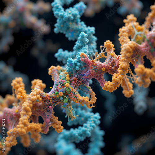
Biological curve protein structure, 3d rendering.
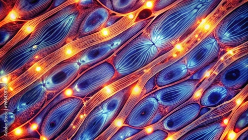
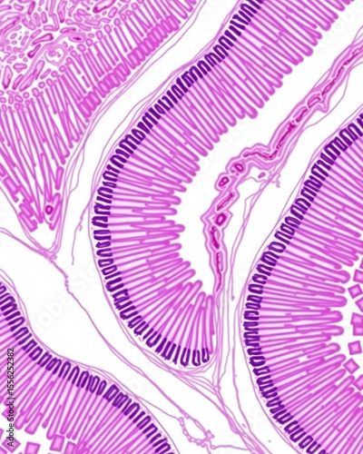
High-contrast microscopic image of smooth muscle fibers exhibiting characteristic spindle-shaped cells, striated texture, and elongated nuclei under brightfield illumination.
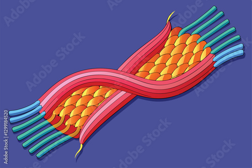
Microscopic view of muscle contraction process, illustrating actin and myosin filaments sliding past each other on a transparent background, ideal for scientific projects
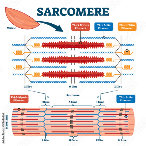
Structures of muscle with fiber, myofibril and sarcomere contraction outline diagram, transparent background.
Aficamten is an investigational, oral, small molecule cardiac myosin inhibitor being developed for the treatment of obstructive hypertrophic cardiomyopathy (HCM)
Protein Cartoon Character
Ouabain, congestive heart failure and supraventricular arrhythmia drug, 3D molecule spinning on Y-axis, 4K
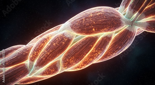
Seethrough muscle texture and sinews, hyperrealistic, superdetailed, clean sharp focus, professional color grading, closeup ,close-up
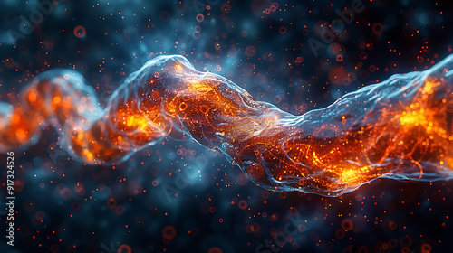
Glowing protein structures a molecular view of actin and myosin filaments. Fluorescent highlights on a dark background. Scientifically accurate representation.
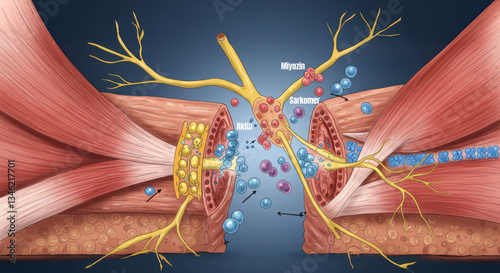
muscle contraction mechanism (Troponin,Tropomyosin,Active site,actin,Myosin head,Actin)
Exotic reddish fruit texture with thick filament surface unique structure on minimal white background showcasing pattern and depth
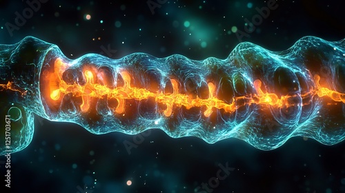
Glowing sarcomere inside a muscle fiber showing actin and myosin filaments interacting during contraction
Muscle Tissue Structure Showing Cells and Fibers Illustration for Science
Skeletal muscle anatomy outline diagram. Structure with anatomical inner layers. Educational medical illustration
Pachysandra terminalis | Dense carpet of Japanese spurge or pachysandra. White upright flowers head with thick filaments above yellowish glossy green alternate leaves
Muscle proteins in action/ Illustration of myosin and actin interacting during muscle contraction
Cross-sectional micrograph of human skeletal muscle fibers under hematoxylin and isomer staining, showing stripes (actin and myosin), peripheral nuclei, and connective tissue. Pink and purple
This visual illustrates how muscles contract through the sliding interaction of myosin and filaments during movement, Muscles contract by sliding myosin and filaments along each other
3D Rendering of Muscle Contraction – Actin & Myosin Filament Interaction
Human cardiac myosin filament 3D molecule, spinning on Y axis, 4K
Detailed illustration of cardiac muscle fibers, highlighting the nuclei and intercalated discs, set against a transparent background
Myosin Filaments Moving Along Actin Fibers in Microscopic View
Visualizing Muscle Fiber Structure with Binding Sites for Medical Illustration
Sarcomere muscular biology scheme illustration, transparent background. Myosin filaments, discs, lines and bands. Myofibril detailed labeled diagram. Sports educational health information.
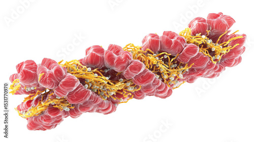
**Cross-Section Of Skeletal Muscle Fiber Showing Actin, Myosin, And Sarcomere Structures. --V 6.1 --S 50 --Raw** - @Darya (Relaxed)
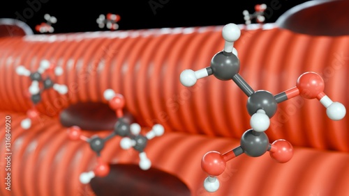
3d rendering of lactic acid molecules and skeletal muscle. The accumulation of lactic acid in muscle tissue during intense physical activity, leading to muscle fatigue and soreness.
Muscle protein structures actin and myosin. Images should showcase proteins with complex details, highlighting their unique shapes and arrangements.
Types of Muscle Response Science Design. Vector Illustration.
Glowing protein structures A molecular view of actin and myosin filaments. Fluorescent highlights on dark background show intricate detail. Scientifically accurate representation.
Cardiac Muscle Tissue - 3d illustration isometric view
Muscle injury
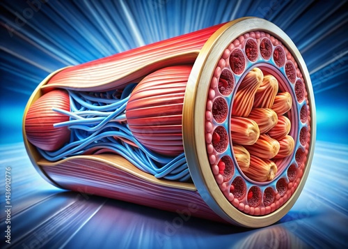
A muscle fiber cross section shows the internal structure and composition
Extreme close-up Muscle fiber contraction. Cellular detail revealed. Bioluminescent highlights. Scientifically accurate. Dark background.
Microscopic View of Muscle Contraction: Neuromuscular Junction
Contracted myofibril made up of myofilaments and proteins.
Adipose tissue human, Soft palate human, Bone human and Striated (skeletal) muscle human under the microscope in Lab isolated with white highlights, png
Skeletal muscle anatomy structure in flat design on white background.
This illustration depicts how muscle fibers contract through the sliding of myosin and actin filaments, Muscles contract by sliding myosin and filaments along each other
3D Rendering of Muscle Contraction – Actin & Myosin Filament Interaction
Muskelfaser
The delicate balance between actin and myosin proteins during cytokinesis allowing for the controlled constriction of the cell
Actomyosin complex, intricate bond between actin and myosin, cellular contraction, 3D illustration
Colorful illustration of human muscle cells with detailed cellular structures, mitochondria, and nuclei, showcasing muscle fibers and tissues in a vibrant, microscopic view.
Detailed Muscle Fiber Structure Illustrating Contraction and Anatomy
From muscle to muscle fiber: tendon muscle muscle fiber.
Semi Realistic Muscle Fiber Diagram Highlighting Sarcomeres and Z-Lines, Cutout Transparent PN
Myosin molecule scientific vector illustration diagram
Types of Muscle Response Science Design. Vector Illustration.
Close-up of Human Muscle Tissue in Contraction Process: Understanding Biology and Movement Function
Blood sample tube and paper order request for troponin I.
A cross section of muscle fibers depicted as tightly packed
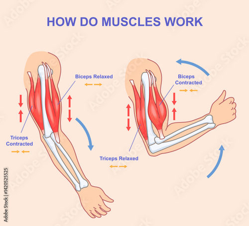
How do muscles work. Educational anatomical diagram showing process of contraction and relaxation of arm muscles. Biceps and triceps movements during sports exercise. Cartoon flat vector illustration
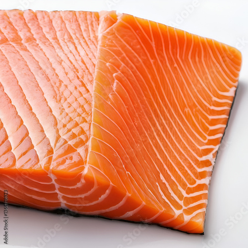
Freshly caught salmon fillet on a white background, showcasing the flaky pink flesh and prominent white muscle striations, highlighting its nutritional value and freshness.
Human Muscle Fiber Anatomy: A Detailed 3D Render
Skeletal muscle anatomy outline diagram. Structure with anatomical inner layers. Labeled educational medical illustration
Close-up of muscle tissue fibers under a microscope, showing the detailed structure of the cells
Glowing sarcomere inside a muscle fiber showing actin and myosin filaments interacting during contraction
Highresolution image depicting process of muscle contraction highlighting the role of actin myosin and the sliding filament theory, muscle contraction actin myosin sliding filament theory
Mavacamten drug molecule. Skeletal formula.
Muscle tissue contracting, showing the sliding filament theory with actin and myosin filaments
Ouabain, cardiac glycoside drug for congestive heart failure and supraventricular arrhythmias, 3D molecule 4K
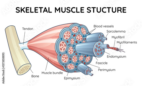
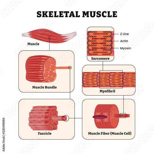
Skeletal muscle structure layers anatomy diagram poster, Labeled with bundle, fascicle, fibers, myofibril and sarcomere sections, z line, actin, myosin. Vector illustration.
Vibrant 3D Illustration of Microscopic Muscle Fiber Contraction
Glowing myosin and actin filaments. A molecular view with fluorescent highlights against a dark background. Scientifically accurate protein structures.
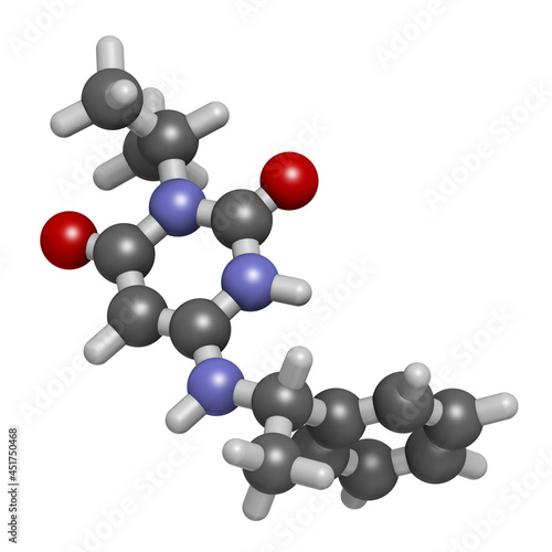
Mavacamten drug molecule, illustration
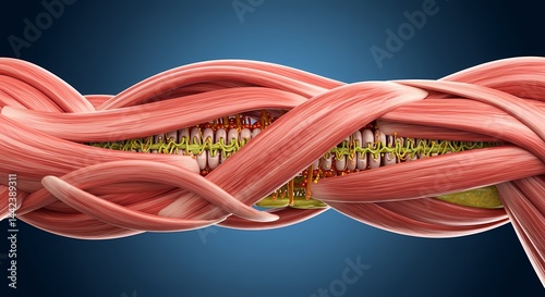
A cut away and zoom on a muscle and its structure: tendon muscle.
Muscle protein structures actin and myosin. Images should showcase proteins with complex details, highlighting their unique shapes and arrangements.
Actomyosin complex, intricate bond between actin and myosin, cellular contraction, 3D illustration
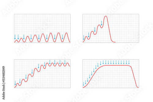
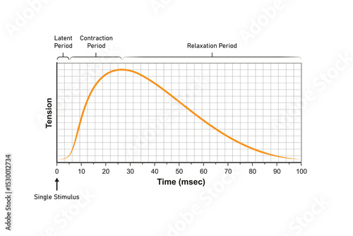
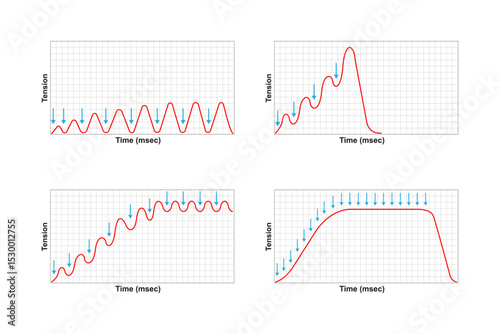
Myogram, Muscle Response of a Single Stimulation Science Design. Vector Illustration.
Microscopic view capturing sarcomere shortening during muscle contraction, highlighting the intricate interaction of actin and myosin filaments against a transparent background
Actomyosin Complex Illustration Isolated on Transparent Background
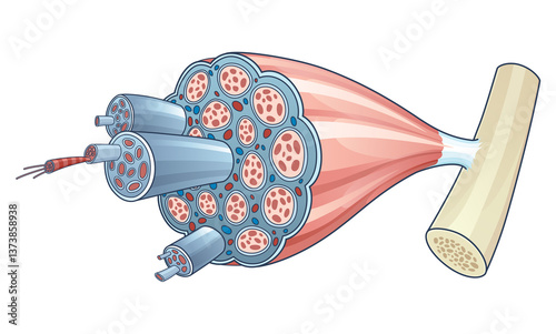
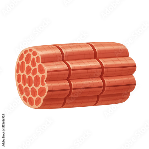
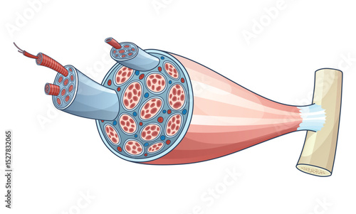
Muscle bundle made up of several largely vascularized muscle fibers.
The human cardiac actin-tropomyosin-myosin complex with ADP-Mg2+
Blood sample tube and paper reqeust order for troponin-I test analysis.
Myosin Filaments Moving Along Actin Fibers in Microscopic View
Skeletal muscle anatomy outline diagram. Structure with anatomical inner layers. Educational medical vector illustration
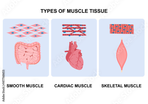
Types of muscle cells include skeletal, smooth, and cardiac muscles. Each muscle specialized for different functions. Medical poster design illustration. Science educational symbol
Glowing sarcomere inside a muscle fiber showing actin and myosin filaments interacting during contraction
Highresolution image depicting process of muscle contraction highlighting the role of actin myosin and the sliding filament theory, muscle contraction actin myosin sliding filament theory
Image of the process of muscle contraction
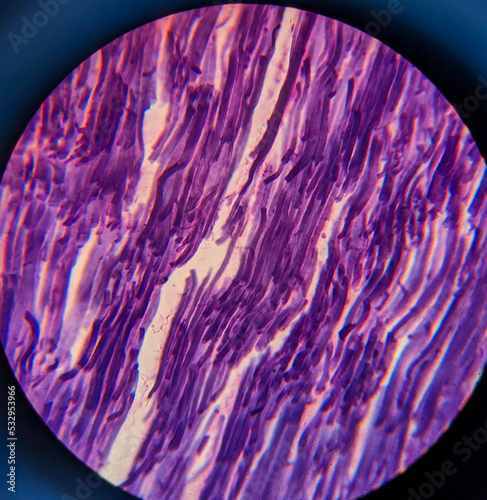
Microscopic View of Striated Muscle Fibers
**Cross-Section Of Skeletal Muscle Fiber Showing Actin, Myosin, And Sarcomere Structures. --V 6.1 --S 50 --Raw** - @Darya (Relaxed)
Biological curve protein structure, 3d rendering.
Cross Section View of Skeletal Muscle Fibers Showing Cell Structure
Actomyosin complex, intricate bond between actin and myosin, cellular contraction, 3D illustration
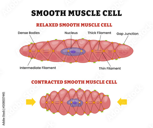
Smooth muscle cells structure anatomy diagram, comparison with relaxed or contracted states and shape differences, labeled gap junction, nucleus, dense bodies, thin and thick filaments.
Mavacamten drug molecule, illustration
Muscle protein structures actin and myosin. Images should showcase proteins with complex details, highlighting their unique shapes and arrangements.
Illustration highlights myofibrils, focusing on the arrangement of thick and thin filaments inside muscle fibers, Myofibril with thin and thick filament,
Types of Muscle Response Science Design. Vector Illustration.
Hyper realistic myosin protein molecular structure biological visualization microscopic scientific vibrant colors futuristic biotechnology cellular function muscle contraction medical research mesh
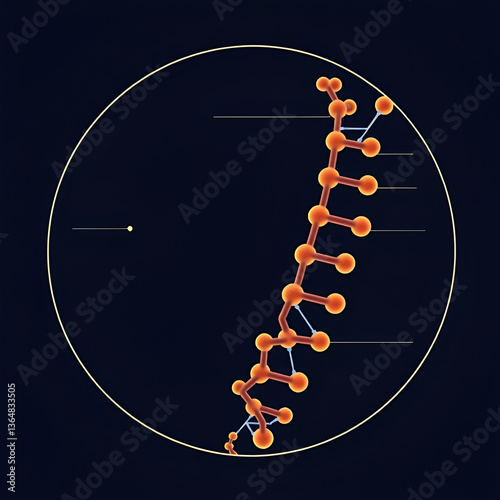
3d rendering of thin filaments, a part of striated muscle that are made up of three proteins: actin, troponin, and . Actin is the main protein component of the thin filament.
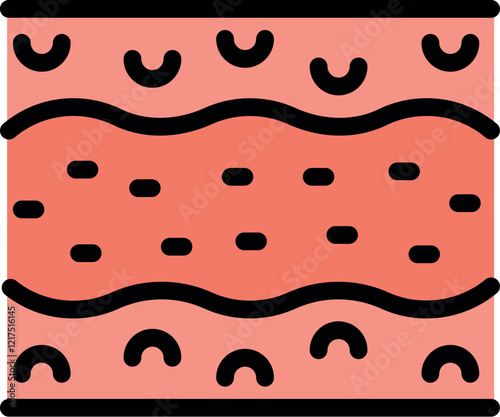
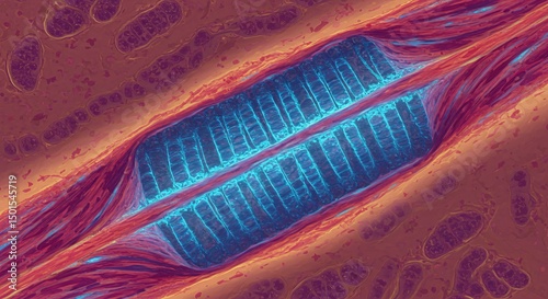
Striated muscle fiber. A band
Muscles: Key tissues that facilitate movement and support posture by contracting and relaxing, providing strength and mobility throughout the body.
photo of tissue under the microscope
Human Muscle Anatomy: Cross-Section Revealing Internal Structure and Active Function
anatomy muscle types muscle groups human body physiology