Login
Sign Up
Member Area
Login
Sign Up
English
Afrikaans formal
Afrikaans informal
Arabic
Azerbaijan
Brazilian
Bulgarian
Catalan
Chinese simplified
Chinese traditional
Croatian
Czech
Danish
Dutch
English
Estonian
Finnish
French
Georgian
German
Greek
Hebrew
Hungarian
Icelandic
Indonesian
Italian
Japanese
Latvian
Lithuanian
Malaysian
Norwegian
Persian
Polish
Portuguese
Romanian
Russian
Serbian
Slovakian
Slovenian
Spanish
Swedish
Thai
Turkish
Ukrainian
Vietnamese
Toggle navigation
VideotonStock
Your Shopping Cart is empty.
Files
Stock
Photo
Video
Special offer
Collections
Packages
Lightboxes
Licenses
Royalty free
Rights managed
Creative
Editorial
Items
Most downloaded
Featured
Most popular
New
Free
Categories
Browse categories
Site info
Site info
About
Support
Privacy Policy
Privacy Policy
FAQ
Contact Us
Contact Us
Terms and Conditions
Buyer Agreement
Seller Agreement
Customers
Users
Credits
Photographers
Boby Richards
0
Boby Richards
0
Videoton1
286
Search
Keywords:
Creator ID:
Type:
Photo
Illustration
Vector
Video
Template
3d
Categories:
Animals
Buildings and Architecture
Business
Drinks
The Environment
States of Mind
Food
Graphic Resources
Hobbies and Leisure
Industry
Landscapes
Lifestyle
People
Plants and Flowers
Culture and Religion
Science
Social Issues
Sports
Technology
Transport
Travel
News
Entertainment
Sport News
Languages:
Brazilan Portuguese - Brazil
English - Canada
French - Canada
Spanish - Mexico
English - United States
English - Belgium
French - Belgium
Dutch - Belgium
Czech - Czech Republic
English - Cyprus
Danish - Danmark
German - Germany
English - Estonia
Spanish - Spain
French - France
French - Marocco
English - Greece
English - Ireland
Italian - Italia
English - Latvia
English - Lithuania
German - Luxembourg
English - Luxembourg
French - Luxembourg
English - Hungary
English - Malta
Dutch - Nederlands
Bokmal Norwegian - Norway
German - Austria
Polish - Poland
Portuguese - Portugal
English - Romania
German - Switzerland
English - Slovenia
Slovakian - Slovakia
French - Switzerland
English - Finland
Swedish- Sweden
Italian - Switzerland
English - United Kingdom
English - Bulgaria
English - Australia
Japanese - Japan
Korean - South Korea
English - New Zealand
Russian - Russia
English - Ukrain
English - Thailand
Orientation:
All
Horizontal
Vertical
square
Color:
Model property release:
All
Yes
No
Duration Video:
All
Up to 10 seconds
Up to 20 seconds
Longer than 30 seconds
Results
(0)
Site
Getty/iStock
Bigstockphoto
Pixabay
Adobe Stock
Relevance
New
Most popular
Most downloaded
Undiscovered
12
24
36
48
96
Auto paging
Menu
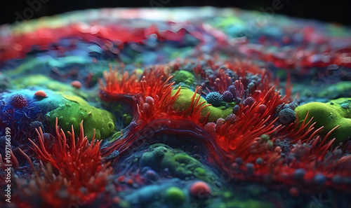
microscopic organism patterns in vibrant colors
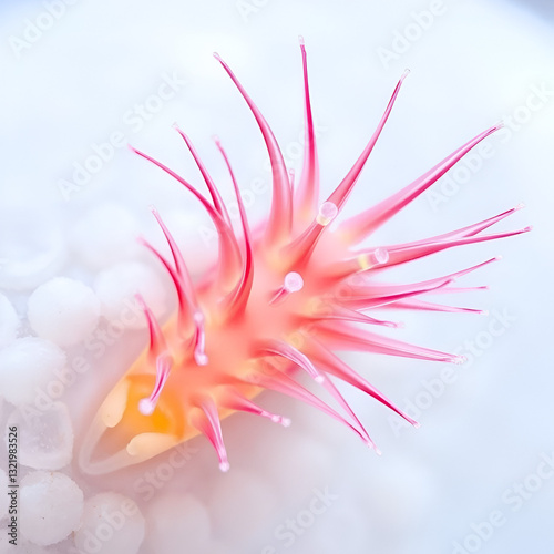
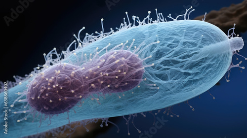
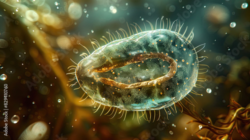
Movement of cilia on a paramecium’s surface, aiding in locomotion and feeding, Animal Digital Art
Amoeba under DIC microscope
microscopic organisms colorful spirals of bacteria and algae
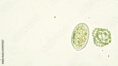
Microscopic image of a paramecium's complex structure.
The Chlamydomonas algae, paramecium ciliates, and bacteria are seen in this microscopy picture, Chlamydomonas microscopy, Chlamydomonas
Ciliates under DIC microscope
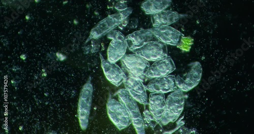
Microscopic view of paramecium
Paramecium Paradise
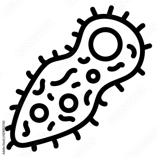
Paramecium Icon
Paramecium caudatum is a genus of unicellular ciliated protozoan and Bacterium under the microscope
Paramecium caudatum under microscope Simplest microorganism. Unicellular protist. Art illustration stylized colorful cell. Cellular structures macro close up. Educational scientific biological project
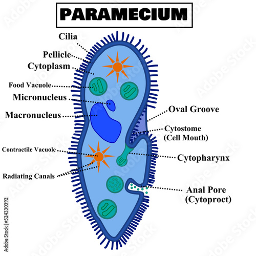
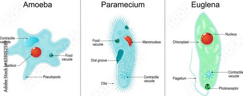
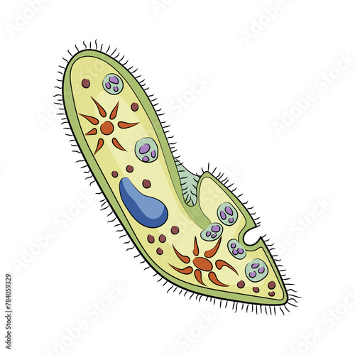
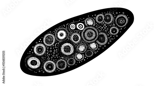
Structure of paramecium caudatum or paramecia with organelles.Ingohraphic for science or biology.Anatomy diagram.Education chart.Cartoon vector illustration isolated on white background.Flat design.
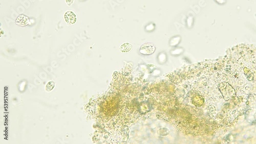
Paramecium and microorganism in pond water under microscope
A highly detailed paramecium with cilia covering its elongated body, isolated on a white background.
Freshwater protozoan and algae under microscope - 100X
Colony of paramecium ciliates move fast in the water seen under optical microscope. 10x objective
Streptococcus, Lactobacillus, Escherichia coli, Proteus Single celled organisms: Chlamydomonas, Paramecium, Cyanobacteria, Amoeba Fungi include yeast, shiitake mushrooms, green mold bacteria, cell wal
Freshwater protozoan and algae under microscope - 100X
Microscopic paramecia release symbiotic green algae into their environment, showcasing a fascinating interaction within aquatic ecosystems and highlighting the complexity of microbial life
Paramecium glyph icon
A microscopic image of a paramecium with a detailed cell structure and multiple cilia.
Amoeba and euglena egg under microscope?
microscopic spiral of algae cells
Ciliate under microscope
Paramecium Paradise
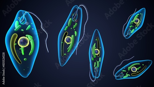
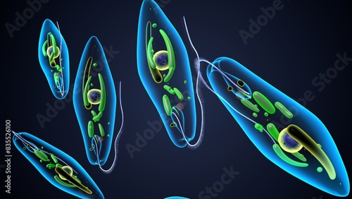
paramecium ciliate, amoeba and Euglena
Streptococcus, Lactobacillus, Escherichia coli, Proteus Single celled organisms: Chlamydomonas, Paramecium, Cyanobacteria, Amoeba Fungi include yeast, shiitake mushrooms, green mold bacteria, cell wal
Detailed depiction of a singlecelled paramecium swimming among debris in a microscopic world
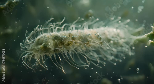
Hyper-realistic Wallpaper Background of Cilia on a Paramecium Waving
Detailed image of a single-celled organism like a paramecium under a microscope highlighting its cilia and nucleus
Amoeba under DIC microscope
Visualization of microorganisms: bright colors, subtle gradations of light.
Microscopic view of Paramecium ciliate with flagella, protozoa, single-celled animal, microscopy
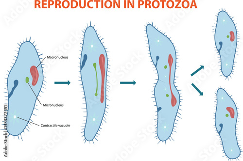
Illustration of protozoa's asexual reproduction diagram
Paramecium multimicronucleatum, LM
Paramecium ciliate float in the water seen under optical microscope. 40x objective
Paramecium multimicronucleatum, LM
The cilia on a paramecium are vividly displayed, showcasing their delicate structure and coloration in a close-up view
Paramecium multimicronucleatum, LM
Paramecium caudatum swimming among plant debris under a microscope.
Microscopic of paramecium and amoeba
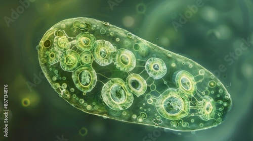
A detailed macro image of a paramecium reveals its clear domeshaped structure and its distinctly curved shape. Its length reaches up to 1 millimeter and is composed
Paramecium and microorganism in pond water under microscope
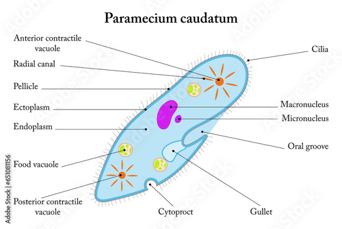
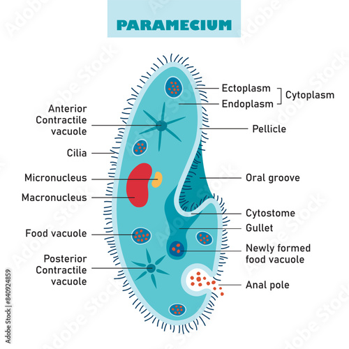
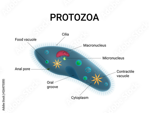
Labelled diagram of Paramecium caudatum.
fresh water ciliate, Paramecium bursaria, with ingested green algae circulating in its cytosol. 450x magnification, phase contrast microscopy
Colorful illustrated paramecium on a white background, highlighting the concept of microorganisms in biology. Vector illustration
Close-up of a paramecium with cilia moving in water, surrounded by pond debris and plant matter.
Microscopic view of a paramecium aurelia with its nucleus, cytoplasm and cilia
microscopic view of protozoa organisms, including amoeba, paramecium and euglena
Hyper-realistic Wallpaper Background of Cilia on a Paramecium
Internal digestive process of a paramecium, showing food vacuoles forming inside the cell, Animal Illustration
Structure of a paramecium
Microscopic view of Chlamydomonas algae, paramecium ciliates, and bacteria , microscopy, Chlamydomonas
Streptococcus, Lactobacillus, Escherichia coli, Proteus Single celled organisms: Chlamydomonas, Paramecium, Cyanobacteria, Amoeba Fungi include yeast, shiitake mushrooms, green mold bacteria, cell wal
Interactive style illustration of paramecium interacting with its environment
Paramecium icon
The Structure Of Paramecium Caudatum
Paramecium glyph icon
Methods of study of Protozoa, Algae and bacteria under the microscope in the biology laboratory.
Detailed microscopic view of a ciliated paramecium in motion
Internal digestive process of a paramecium, showing food vacuoles forming inside the cell, animal illustration
Paramecium bursaria with symbiotic algea differential interference contrast DIC focus to macronucleus oral apparatus mouth field symbiotic algea extrusomes
Paramecium under microscope
The cilia on a paramecium are vividly displayed, showcasing their delicate structure and coloration in a close-up view
euglena reproduction anatomy. 3d render
Creative Hand Drawn vector silhouette illustration of an infusoria slipper (Paramecium) showing internal organelles, a unique single-celled microorganism from the microscopic world.
Icon of Paramecium Caudatum
Paramecium Closeup
microscopic view of protozoa organisms, including amoeba, paramecium and euglena
Protozoa Cell Structure and Anatomy
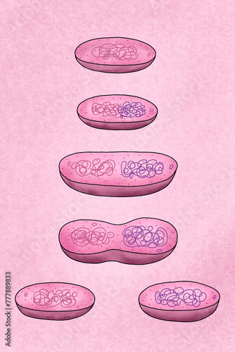
Binary fission of bacteria
Microscopic of paramecium and amoeba
Close-up of a paramecium with cilia moving in water, surrounded by pond debris and plant matter.
microscopic swirls of aquatic organisms in vibrant hues
Internal organelles of a paramecium, showing contractile vacuole and micronucleus, Animal Vector Graphic
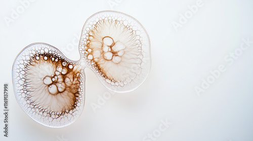
Microscopic paramecium division stage, cilia active, floating mid-split on white background
Ciliates under DIC microscope
Internal organelles of a paramecium, showing contractile vacuole and micronucleus, Animal Vector Graphic
Paramecium Icon
Methods of study of Protozoa, Algae and bacteria under the microscope in the biology laboratory.
Detailed depiction of a singlecelled paramecium swimming among debris in a microscopic world
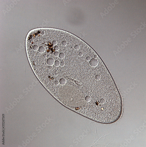
Microscopic view of Paramecium caudatum's asymmetrical body, cilia, and macronucleus, with distinctive caudal tail, amidst blurred aquatic background.
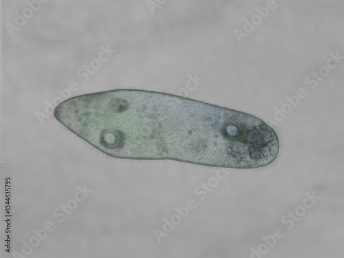
Paramecium caudatum
Ciliates under DIC microscope
Microscopic view of paramecium
euglena reproduction anatomy. 3d render
A highly detailed paramecium with cilia covering its elongated body, isolated on a white background.
Microscopic Universe: A vibrant paramecium dominates, cilia aglow, in a bustling microscopic ecosystem.
Binary fission
Single celled Paramecium's hunting in teal colored water
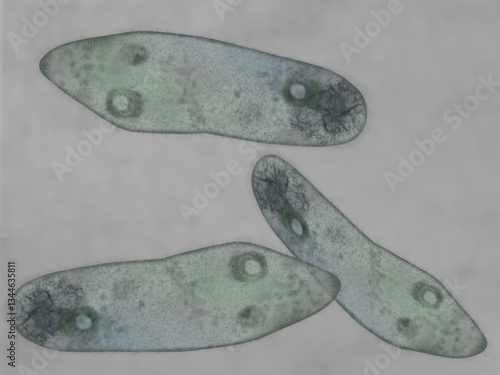
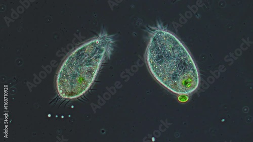
Two paramecium interacting with each other, enlarged protozoa under a microscope.
amoeba
Close-up of a paramecium with cilia moving in water, against a pond backdrop with plant matter.
microscopic view of protozoa organisms, including amoeba, paramecium and euglena
a closeup or macro image of paramecium caudatum on white background
Microscopic view of a paramecium, a single-celled organism
Internal digestive process of a paramecium, showing food vacuoles forming inside the cell, animal illustration
animal conjugation of Paramecium caudatum
Educational game: assembling Paramecium caudatum from ready-made components in form of stickers. Paramecium caudatum structure with titles