Login
Sign Up
Member Area
Login
Sign Up
English
Afrikaans formal
Afrikaans informal
Arabic
Azerbaijan
Brazilian
Bulgarian
Catalan
Chinese simplified
Chinese traditional
Croatian
Czech
Danish
Dutch
English
Estonian
Finnish
French
Georgian
German
Greek
Hebrew
Hungarian
Icelandic
Indonesian
Italian
Japanese
Latvian
Lithuanian
Malaysian
Norwegian
Persian
Polish
Portuguese
Romanian
Russian
Serbian
Slovakian
Slovenian
Spanish
Swedish
Thai
Turkish
Ukrainian
Vietnamese
Toggle navigation
VideotonStock
Your Shopping Cart is empty.
Files
Stock
Photo
Video
Special offer
Collections
Packages
Lightboxes
Licenses
Royalty free
Rights managed
Creative
Editorial
Items
Most downloaded
Featured
Most popular
New
Free
Categories
Browse categories
Site info
Site info
About
Support
Privacy Policy
Privacy Policy
FAQ
Contact Us
Contact Us
Terms and Conditions
Buyer Agreement
Seller Agreement
Customers
Users
Credits
Photographers
Boby Richards
0
Boby Richards
0
Videoton1
286
Search
Keywords:
Creator ID:
Type:
Photo
Illustration
Vector
Video
Template
3d
Categories:
Animals
Buildings and Architecture
Business
Drinks
The Environment
States of Mind
Food
Graphic Resources
Hobbies and Leisure
Industry
Landscapes
Lifestyle
People
Plants and Flowers
Culture and Religion
Science
Social Issues
Sports
Technology
Transport
Travel
News
Entertainment
Sport News
Languages:
Brazilan Portuguese - Brazil
English - Canada
French - Canada
Spanish - Mexico
English - United States
English - Belgium
French - Belgium
Dutch - Belgium
Czech - Czech Republic
English - Cyprus
Danish - Danmark
German - Germany
English - Estonia
Spanish - Spain
French - France
French - Marocco
English - Greece
English - Ireland
Italian - Italia
English - Latvia
English - Lithuania
German - Luxembourg
English - Luxembourg
French - Luxembourg
English - Hungary
English - Malta
Dutch - Nederlands
Bokmal Norwegian - Norway
German - Austria
Polish - Poland
Portuguese - Portugal
English - Romania
German - Switzerland
English - Slovenia
Slovakian - Slovakia
French - Switzerland
English - Finland
Swedish- Sweden
Italian - Switzerland
English - United Kingdom
English - Bulgaria
English - Australia
Japanese - Japan
Korean - South Korea
English - New Zealand
Russian - Russia
English - Ukrain
English - Thailand
Orientation:
All
Horizontal
Vertical
square
Color:
Model property release:
All
Yes
No
Duration Video:
All
Up to 10 seconds
Up to 20 seconds
Longer than 30 seconds
Results
(0)
Site
Getty/iStock
Bigstockphoto
Pixabay
Adobe Stock
Relevance
New
Most popular
Most downloaded
Undiscovered
12
24
36
48
96
Auto paging
Menu
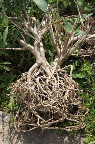
Racines nues d'un petit arbre
緑の中に佇む石製水盤と光沢のある金属製蛇口
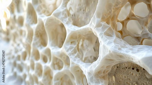
A piece of bread with a lot of holes in it, macro shot
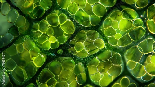
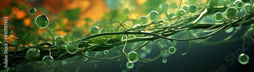
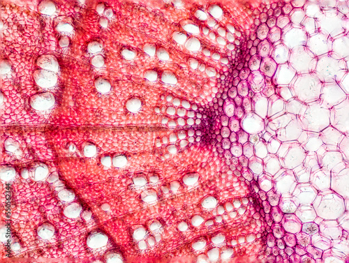
Microscopic view of a plant's parenchyma cells reveals internal structures and vibrant green coloring.
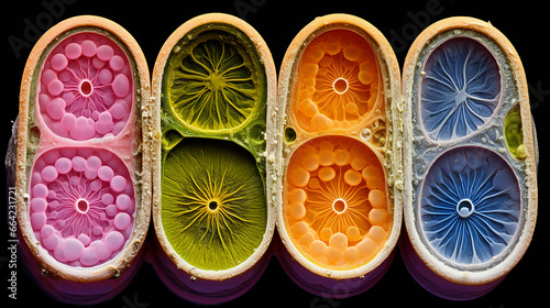
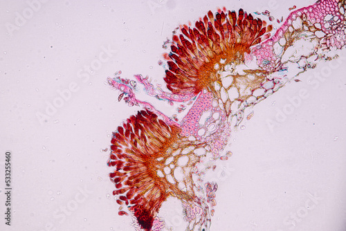
Flower Ovary Cut Open To Show Ovules
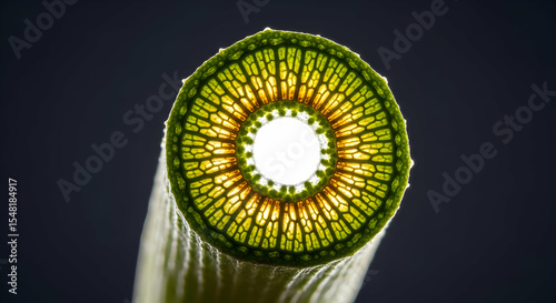
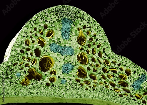
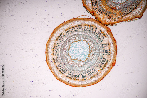
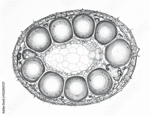
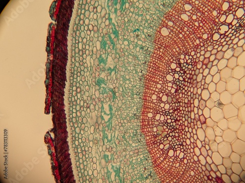
Plant Stem Cross Section Showing Vascular Bundle and Parenchyma Cells Under Microscope
Detailed Cross Section of a Leaf with Complex Cellular Structures
Kiwi w całości i przekrojone, ukazujące szczegóły struktury miąższu
Fern gametophyte under microscope. Fresh sample without staining. Selective focus image
The study of plant tissues under the microscope in the laboratory.
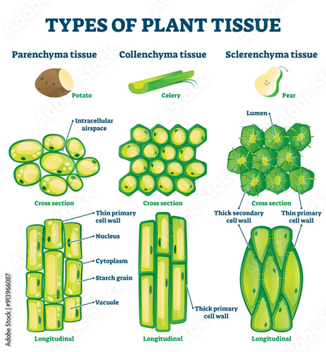
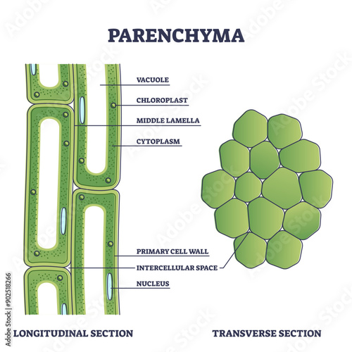
Types of plant tissue illustration, transparent background.Labeled educational structure scheme.Biological closeup with cross section and longitudinal views.
Green potted plant in a white pot resting on a white desk.
一万円札とブロックの上に書かれた実質賃金 経済
Diffused Sunlight Illuminating Tree Foliage and Woody Stems
Water lily stem, SEM
An image of a cross section of a plant stem reveals vibrant green parenchyma cells densely packed and full of chloroplasts responsible
Intricate Veins Of A Teal Leaf Against Soft Blue Gradient Backdrop
The Story of a Tree: Understanding Conservation Through Growth Rings
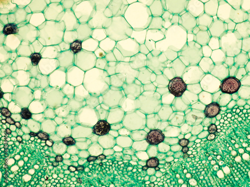
Stem of cotton x.s. details under biological optical misroscope
Cell structure flower, View of chromoplast showing in plant cells under the microscope for classroom education.
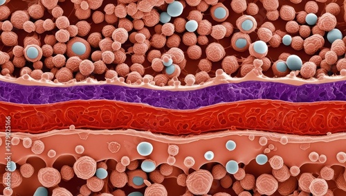
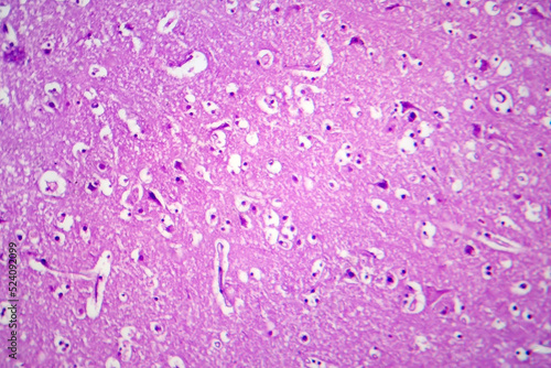
Histopathology of Japanese encephalitis, light micrograph
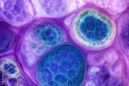
Close-up of a cellular pattern, illustrating the intricate beauty of biological textures in a macro scientific context
Microscopic Plant Tissue Reveals Complex Cell Structure Under Bright Field Illumination
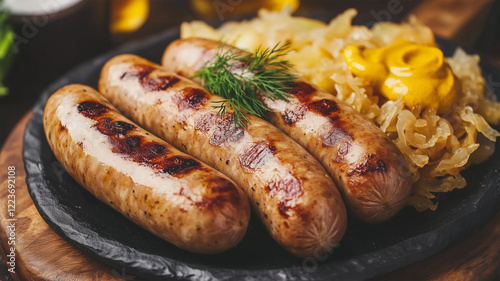
石製プレートに盛られたブラートヴルストとザワークラウトの風味豊かなドイツ料理
Microscopic view of a plant's parenchyma cells reveals internal structures and vibrant green coloring.
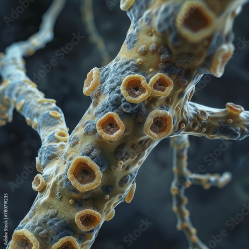
Subepidermal Parenchyma Cells in Young Stem
3D rendered image of parenchyma cells within a leaf, detailed and vibrant, emphasizing their central role in the plants life support system
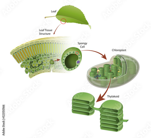
Diagram of a leaf structure
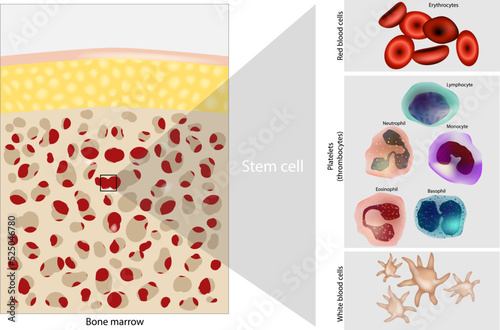
Bone marrow Stem cell. Platelets, Red and White blood cells. Diagram
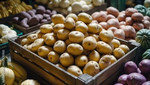
Close up of potato in wooden crate. Fresh potato in the market.
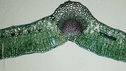
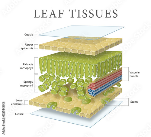
Microbotany A cross section of a leaf. Epidermis, mesophyll, parenchyma, stomata, network of veins. Vegetative organ of a plant whose main functions are photosynthesis, gas exchange, and transpiratio
Exploring the intricate cellular arrangement within a plant stem uncovers its robust structure essential for support and nutrient transport.
Cross sections of plant stem under microscope view show Structure of Parenchyma Cells.
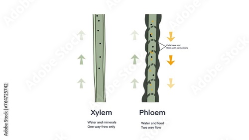
Xylem and phloem water and minerals transportation system outline diagram, Scientific Designing of Xylem And Phloem Scheme, Nutrient And Mineral Transportation, plants transport nutrient and water
Diffused Sunlight Illuminating Tree Foliage and Woody Stems
The Story of a Tree: Understanding Conservation Through Growth Rings
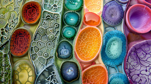
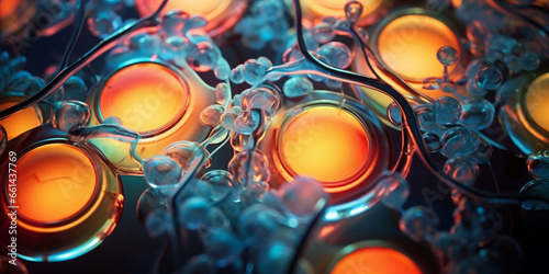
A colorful abstract painting with many small circles of different colors
Cucurbita pepo stem
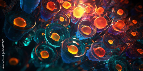
cells brightly colored cells glowing under the microscope
Parenchyma as ground filler tissue for plant stem and roots outline diagram, transparent background. Labeled educational microscopic explanation.
fern root
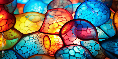
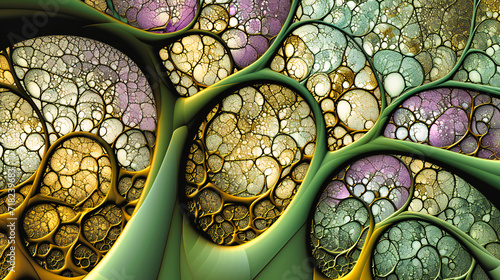
Colorful Cellular Pattern, Abstract Science Background, Artistic Fractal Design, Biological Inspiration
Microscopic View of a Plant Stem Under a Microscope, Green, Blue, Red, Cells, microscopy , botany
実質利回りと書かれたブロック、電卓、スマホ、建物の模型
石製プレートに盛られたブラートヴルストとザワークラウトの風味豊かなドイツ料理
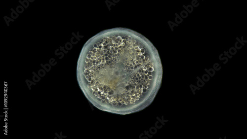
Coral larva under a microscope, order Actiniaria, phylum Cnidaria, adult stage, will soon sink to the bottom and turn into a coral polyp. Atlantic Ocean
Parenchyma Cell Arrangement in Rhizome Cortex Anatomy
Nature study image of a sectioned fruit, emphasizing parenchyma cells crucial role in texture and nutrient storage in edible plants
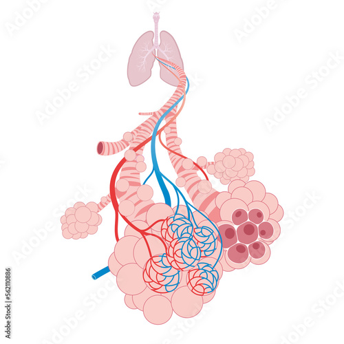
Pulmonary alveoli, trachea, and bronchiole in the lungs
Plant tissue Structure, section (tissue) of stem plant tissue under a light microscope.
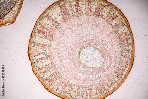
Detailed Cross-Section of a Plant Stem Revealing Cellular Structure and Vascular Bundles
Microscopic image of a stem of Gossypium hirsutum.
Stoma or stomate is a pore found in the epidermis of leaves, stems, and other organs. Opening and closing of stoma
Cały ananas z zielonymi liśćmi i pokrojone kawałki soczystego miąższu na białym tle
Microscopic view, woody dicot stem cross-section, plant cells, lignin, cell structures clearly visible
Diffused Sunlight Illuminating Tree Foliage and Woody Stems
Human kidney cross section anatomy
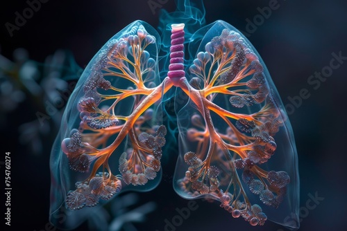
The mesmerizing complexity of the lungs
cells brightly colored cells glowing under the microscope
fern petiole cross section under the microscope - optical microscope x32 magnification
Futuristic medical lung anatomy concept in low-poly style with geometric background
透明な背景のミニマリストな木製フレームと白いクッションを備えたエレガントな椅子、モダンなインテリアデザイン
Microscopic Cross Section of a Plant Stem Showing Vascular Bundles and Cellular Structure
実質と書かれたブロックを掴む手
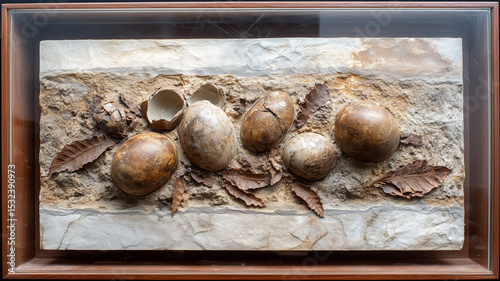
平面展示された恐竜卵巣化石と地層の詳細
Leech Hirudinea close up, class Clitellata. Found on the shrimp Sclerocrangon boreas. Species not determined. White sea
Rhizome Parenchyma Cells Storing Sugars and Oils
Storage Parenchyma Cells with Dense Starch Granules
Leaf tissues and cell layers
Plant tissue Structure, section (tissue) of stem plant tissue under a light microscope.
Microscopic view of a plant's parenchyma cells reveals internal structures and vibrant green coloring.
detailed plant cell structure model white background biological diagram educational resource science art, cytoplasm, chloroplast, vacuole, nucleus
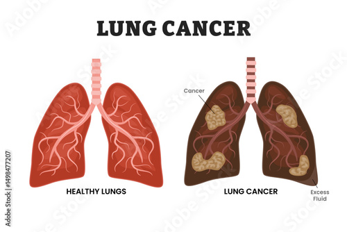
Healthy Lungs vs Lung Cancer or bronchogenic carcinoma,, educational medical poster design healthcare scheme, human lung disease internal organs. Labeled excess fluid, malignant tumor, parenchyma.
Soczyste przekrojone kiwi w zbliżeniu, widoczna faktura miąższu
Cross-section, woody dicot stem, plant cells, microscopic image, stained cells, clear cell walls, cellular organization
photo of section of plant tissue under the microscope
Composite image showing different types of plant cells (epidermal, parenchyma, xylem, phloem) with distinct features and functions
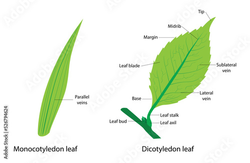
illustration of Plant Biology and botany, Characteristics of monocots and cotyledons, Difference Between Cotyledon Leaf and Cotyledon Leaf
cells brightly colored cells glowing under the microscope
rose of sharon stem transversal section under the microscope - optical microscope x100 magnification
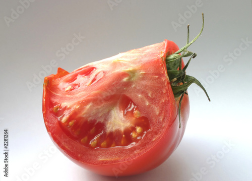
tomato details
透明な背景にベージュ色の革のドレスシューズのペアのクローズアップファッションとエレガントなスタイルと男性のためのデザイナークラシック
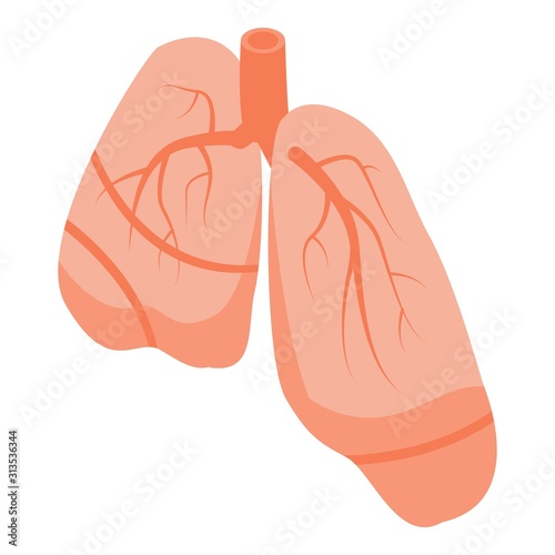
Human lungs icon. Isometric of human lungs vector icon for web design isolated on white background
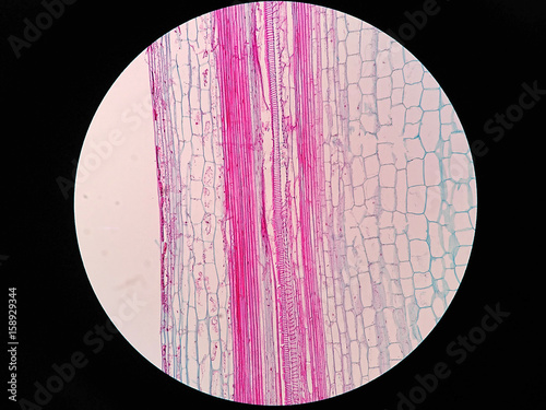
Plant stem, long section under microscope view
平面展示された恐竜卵巣化石と地層の詳細
Cross-section, woody dicot stem, plant cells, microscopic image, stained cells, clear cell walls, cellular organization
Turgor Pressure Effects in Parenchyma Cells
Close up Plant epidermis with stomata or Leaf Epidermis (Stomata) under microscope.
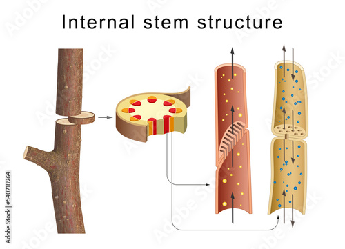
Internal anatomy of the tree stem
Plant tissue Structure, section (tissue) of stem plant tissue under a light microscope.
Microscopic photography of Steam of wood dicotyledon, cross section.
Bacterial pneumonia animation.
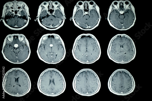
MRI scan of patient brain
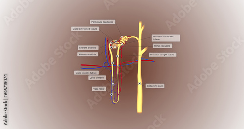
The function of the nephron is to convert blood to urine and consists of the tubular system and the renal corpuscle.
Microscopic photography of the biological objects
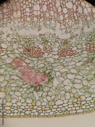
Microscopic Views of Different Plant Tissues in Vivid Detail
実質利回りと書かれたブロック、電卓、不動産の模型
Sieve Tube Food Transportation vintage illustration.
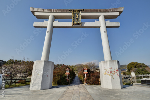
長岡天満宮の鳥居
Microscopic View of Plant Cells in a Leaf Transverse Section Showcasing the Cell Structure and Cellular Arrangement