Login
Sign Up
Member Area
Login
Sign Up
English
Afrikaans formal
Afrikaans informal
Arabic
Azerbaijan
Brazilian
Bulgarian
Catalan
Chinese simplified
Chinese traditional
Croatian
Czech
Danish
Dutch
English
Estonian
Finnish
French
Georgian
German
Greek
Hebrew
Hungarian
Icelandic
Indonesian
Italian
Japanese
Latvian
Lithuanian
Malaysian
Norwegian
Persian
Polish
Portuguese
Romanian
Russian
Serbian
Slovakian
Slovenian
Spanish
Swedish
Thai
Turkish
Ukrainian
Vietnamese
Toggle navigation
VideotonStock
Your Shopping Cart is empty.
Files
Stock
Photo
Video
Special offer
Collections
Packages
Lightboxes
Licenses
Royalty free
Rights managed
Creative
Editorial
Items
Most downloaded
Featured
Most popular
New
Free
Categories
Browse categories
Site info
Site info
About
Support
Privacy Policy
Privacy Policy
FAQ
Contact Us
Contact Us
Terms and Conditions
Buyer Agreement
Seller Agreement
Customers
Users
Credits
Photographers
Boby Richards
0
Boby Richards
0
Videoton1
286
Search
Keywords:
Creator ID:
Type:
Photo
Illustration
Vector
Video
Template
3d
Categories:
Animals
Buildings and Architecture
Business
Drinks
The Environment
States of Mind
Food
Graphic Resources
Hobbies and Leisure
Industry
Landscapes
Lifestyle
People
Plants and Flowers
Culture and Religion
Science
Social Issues
Sports
Technology
Transport
Travel
News
Entertainment
Sport News
Languages:
Brazilan Portuguese - Brazil
English - Canada
French - Canada
Spanish - Mexico
English - United States
English - Belgium
French - Belgium
Dutch - Belgium
Czech - Czech Republic
English - Cyprus
Danish - Danmark
German - Germany
English - Estonia
Spanish - Spain
French - France
French - Marocco
English - Greece
English - Ireland
Italian - Italia
English - Latvia
English - Lithuania
German - Luxembourg
English - Luxembourg
French - Luxembourg
English - Hungary
English - Malta
Dutch - Nederlands
Bokmal Norwegian - Norway
German - Austria
Polish - Poland
Portuguese - Portugal
English - Romania
German - Switzerland
English - Slovenia
Slovakian - Slovakia
French - Switzerland
English - Finland
Swedish- Sweden
Italian - Switzerland
English - United Kingdom
English - Bulgaria
English - Australia
Japanese - Japan
Korean - South Korea
English - New Zealand
Russian - Russia
English - Ukrain
English - Thailand
Orientation:
All
Horizontal
Vertical
square
Color:
Model property release:
All
Yes
No
Duration Video:
All
Up to 10 seconds
Up to 20 seconds
Longer than 30 seconds
Results
(0)
Site
Getty/iStock
Bigstockphoto
Pixabay
Adobe Stock
Relevance
New
Most popular
Most downloaded
Undiscovered
12
24
36
48
96
Auto paging
Menu
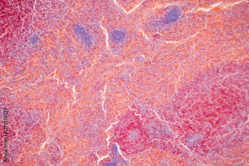
Showing Light micrograph of the Adrenal gland, Spleen human and Other human tissues under the microscope for education in the laboratory.
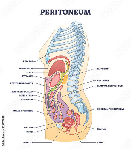
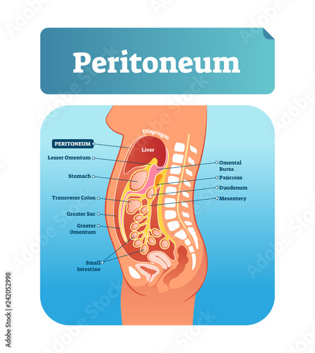
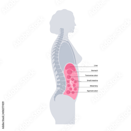
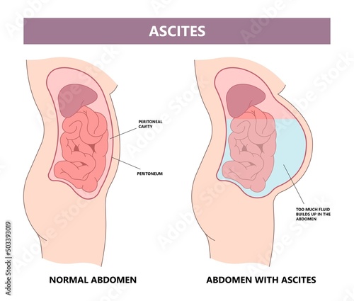
Peritoneum membrane as smooth muscle tissue in abdominopelvic cavity outline diagram. Labeled educational scheme with inner stomach organs from side view vector illustration. Human body organ anatomy.
Macro Photography: Simple Squamous Epithelium Cell Structure - Microscopic View for Education
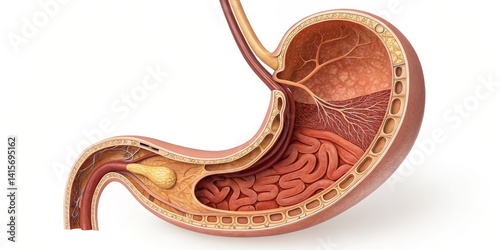
Human Stomach Anatomy 3D Cross-Section Illustration, Digestive System, Internal Organs Anatomy,Digestive System
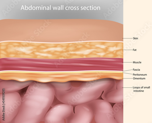
Abdominal wall Layers, muscles and fascia. Cross-section Anatomy Abdominal wall. Medical Illustration
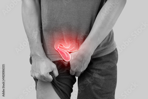
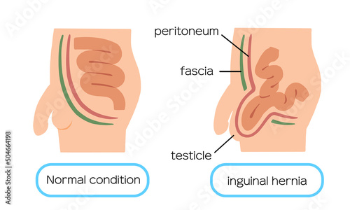
Illustration depicts hernia symptoms in a man's body. Common signs of a hernia are highlighted.
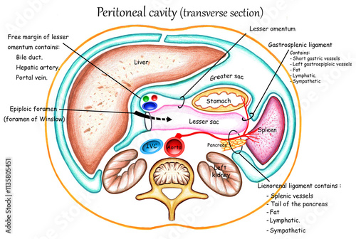
Peritoneal cavity (transverse section)
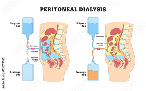
how peritoneal dialysis works, including placement of catheter, dialysate inflow, and drainage from the peritoneal cavity diagram hand drawn schematic vector illustration. Medical science educational
World Ovarian Cancer is observed every year on May 8.It is related areas of the fallopian tubes and the peritoneum
An anatomical diagram depicts peritonitis, emphasizing its inflammation, symptoms, and treatment strategies in a clear and informative manner.
Stages of ovarian cancer vector illustration
The diaphragm separates the chest cavity from the abdomen, where the stomach, liver, and kidneys are nestled among layers of abdominal muscles.
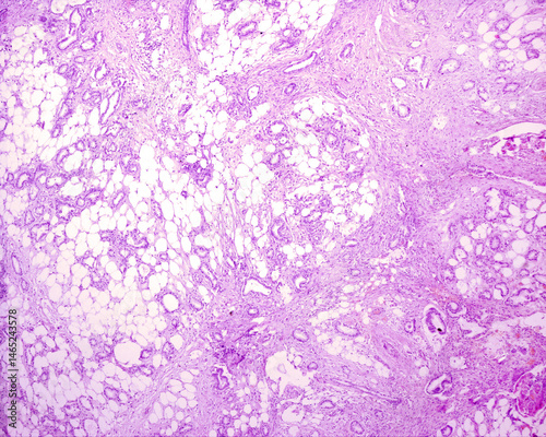
Undifferentiated adenocarcinoma. Metastatic nodule
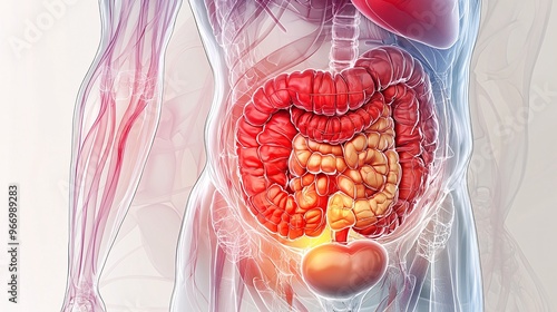
A colorful, highly detailed illustration of the human digestive system, featuring labeled organs and structures,
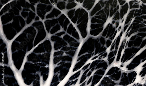
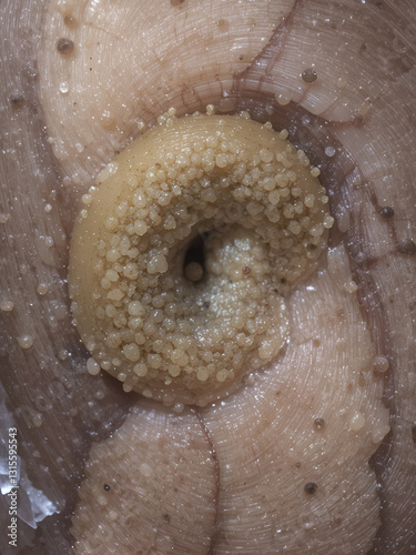
Close-up of a membrane that covers internal organs - Visceral peritoneum
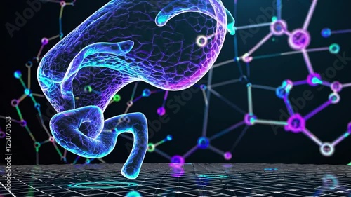
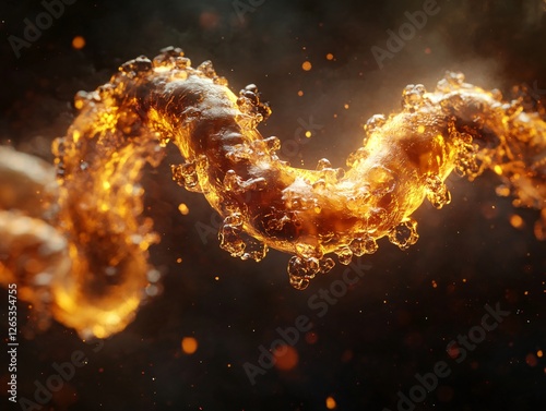
3D-rendered human peritoneum floating in a dark, futuristic space, illuminated by intricate glowing protective and vascular pathways in shades of neon blue, electric purple, and pulsating cyan.
Visualization of a crosssection of the intestines, showing the layers of tissue and muscle, Crosssection of intestines, Anatomical and precise
Glowing Green Abdominal Organs 3D Medical Visualization
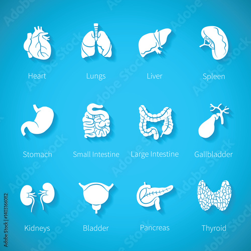
Vector icon set of human internal organs in flat style
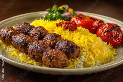
Classic dishes of beef Kofta kebabs and tarb kufta made of minced meat wrapped in lamb fat and grilled served with yellow Basmati rice
Showing Light micrograph of the Adrenal gland, Spleen human and Other human tissues under the microscope for education in the laboratory.
Illuminated 3D illustration of the human stomach's intricate anatomy, showcasing its layered walls, gastric folds, and digestive enzymes amidst a vibrant blue and pink color scheme.
Abdominal wall Layers, muscles and fascia. Cross-section Anatomy Abdominal wall. Medical Illustration
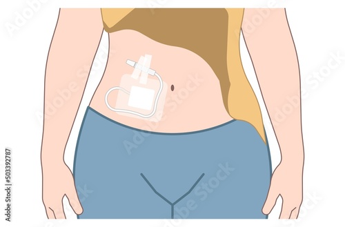
Peritoneal Dialysis Before After Home Care with Dialysate and Drainage Bag, Educational Medical Poster Design Healthcare Scheme, Labeled Catheter, Peritoneum, Connector, Solution, Cavity.
The seven layers of the abdominal wall that must be cut through in order to perform a cesarean section.
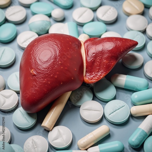
An illustration displaying human liver anatomy against a backdrop of medicine pills
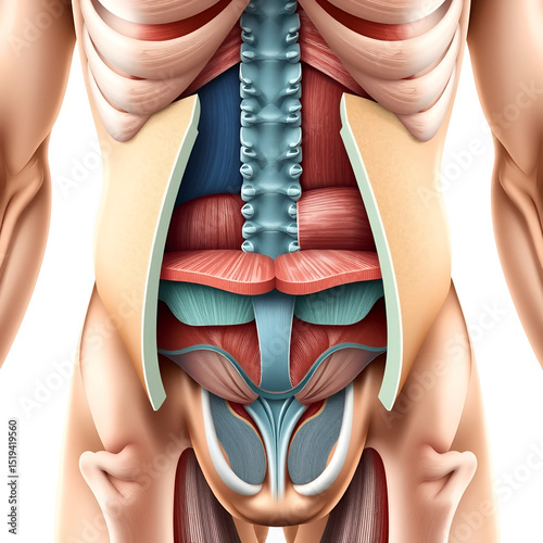
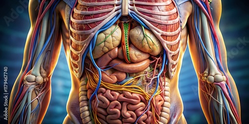
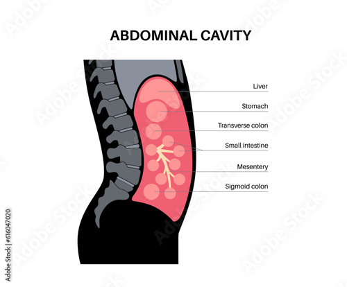
Detailed Medical Diagram of Human Abdominal Cavity Organs
how peritoneal dialysis works, including placement of catheter, dialysate inflow, and drainage from the peritoneal cavity diagram hand drawn schematic raster illustration. Medical science educational
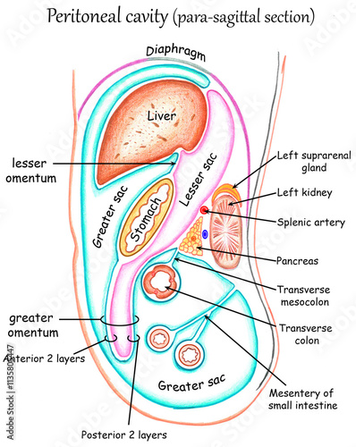
Peritoneal cavity (para-sagittal section)
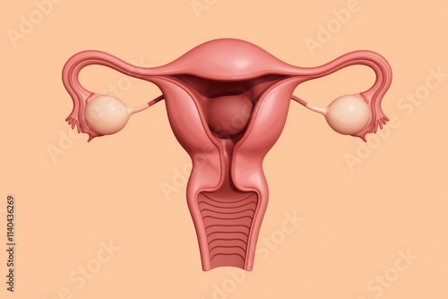
3D model of the female reproductive system showing uterus fallopian tubes and ovaries.
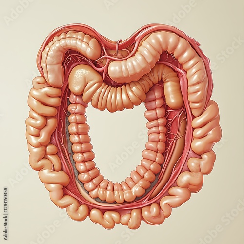
Human Digestive System Anatomy Organs
This image showcases the internal anatomy of the abdominal cavity, featuring the stomach's pear-shaped structure and
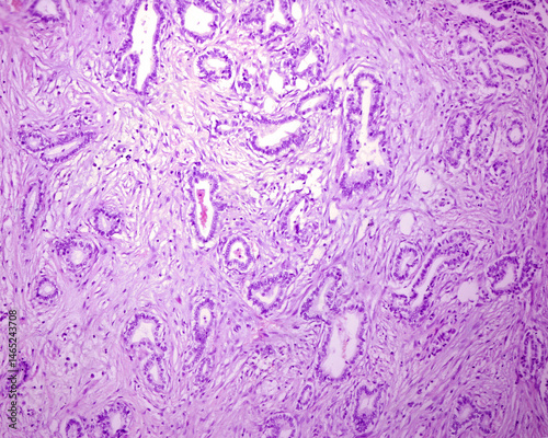
Human uterine tube wall
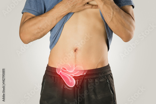
Hernia symptoms shown in body illustration.
Minimalist Abdominal Digestive System Anatomy Illustration
Undifferentiated adenocarcinoma. Metastatic nodule
Intestine Anatomy, 3D Render
Human digestive system isolated,3D rendering
Vector icon set of human internal organs in flat style with shadow
Classic beef kofta kebab and tarb kufta minced meat wrapped in lamb fat grilled and served with yellow Basmati rice
Showing Light micrograph of the Adrenal gland, Spleen human and Other human tissues under the microscope for education in the laboratory.
Detailed illustration of the abdominal muscle anatomy, showcasing the rectus abdominis, external obliques, and internal
Abdominal wall cross section Anatomy. Abdominal wall layers Medical Illustration
Paraovarian cyst
A holographic display of the human digestive system, showcasing the transparent layers of the stomach, intestines, and related organs, with glowing lines highlighting the digestive tract
An illustration displaying human liver anatomy against a backdrop of medicine pills
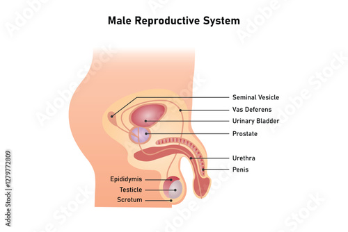
Male Reproductive System Science Design. Vector Illustration.
Panoramic Medical Anatomy Poster: Stomach & Viscera - Detailed Illustration
Peritoneal covering of the stomach
External urethral sphincter controlling urine flow
Stunning Detailed Medical Illustration of Peritonealizing in the Human Abdominal Cavity Anatomy
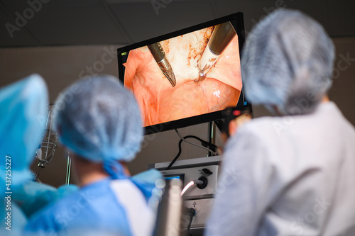
Monitor depicting endoscopic surger
Adenocarcinoma. Metastatic nodule
Most common sites of Endometriosis Unlabeled diagram Side view Transparent PNG
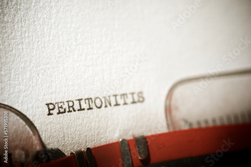
Peritonitis concept view
Peritoneum vector illustration. Labeled medical membrane location scheme.
dialysate cleansing home care tube fluid drain bag waste High blood cysts center manual port End Stage renal ESRD Infusion Diffusion fatty liver tap cancer B C Use pain cavity portal peritoneocentesis
Sigmoid
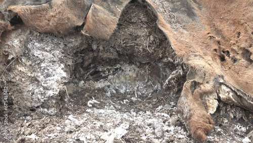
The skin-eaters gnawed holes in the cow's hide. Dogs and foxes ate the peritoneum
Classic beef Kofta kebab and tarb kufta minced meat in lamb fat grilled served with yellow Basmati rice
Showing Light micrograph of the Adrenal gland, Spleen human and Other human tissues under the microscope for education in the laboratory.
Superior Mesenteric Artery and Its Branches.3d rendering
Illustrated diagram of the human abdomen showing internal organs including kidneys, liver, stomach, small intestine,
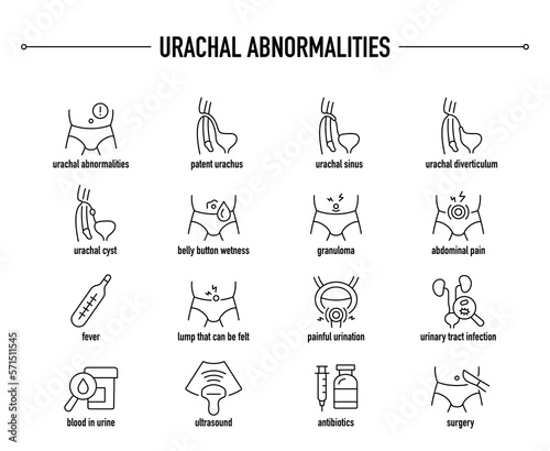
Urachal Abnormalities symptoms, diagnostic and treatment vector icon set. Line editable medical icons.
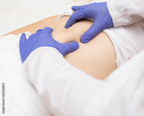
Doctor surgeon examines the stomach of a girl patient for the presence of umbilical hernia. Abdominal wall disease concept with umbilical hernia, reinforced hernia
GYNECOLOGICAL SURGERY
An illustration displaying human liver anatomy against a backdrop of medicine pills
Abdominal cavity anatomy
Abdominals
Peritoneal dialysis
Chef wraps the roll of the peritoneum with garlic.
Monitor depicting endoscopic surgery. Endoscopic camera
Monitor depicting endoscopic surger
Adenocarcinoma. Metastatic nodule
Ultra-Detailed Human Gastrointestinal Tract Icon for Medical Illustrators
Peritonitis concept view
Male illness inguinal hernia illustration
dialysate cleansing home care tube fluid drain bag waste High blood cysts center manual port End Stage renal ESRD Infusion Diffusion fatty liver tap cancer B C Use pain cavity portal peritoneocentesis
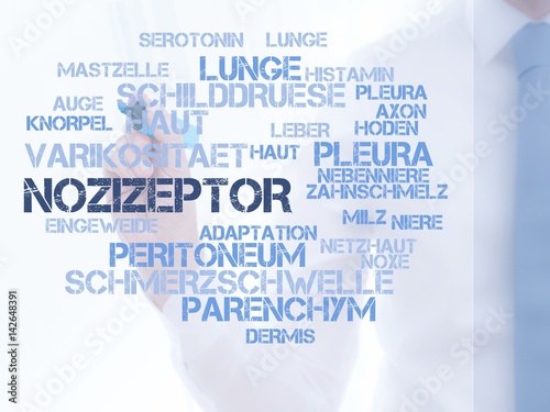
Nozizeptor
Hologram screen 3d of the small intestine of the human body - loop
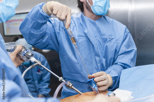
Close up surgical operation using laparoscopic equipment in modern clinic
Showing Light micrograph of the Adrenal gland, Spleen human and Other human tissues under the microscope for education in the laboratory.
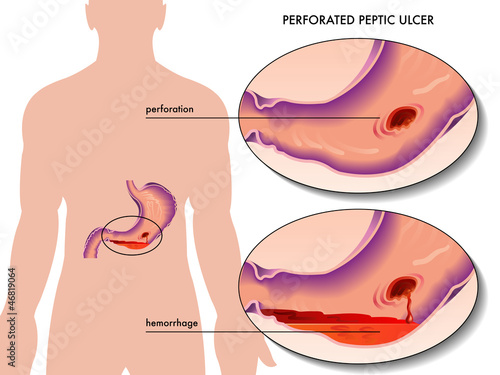
ulcera peptica perforata
Medical anatomy poster stomach viscera concept
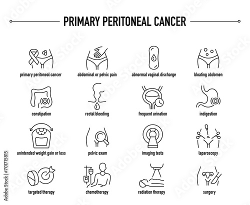
Primary Peritoneal Cancer symptoms, diagnostic and treatment vector icons. Line editable medical icons.
The abdominal viscera can be divided anatomically by their relationship to the peritoneum
Bauchfell. Arzt im Kittel hält Sprechblase hoch. Das Wort Bauchfell steht im Schild. Symbol für Krankheit, Gesundheit, Medizin
Abdominal cavity anatomy
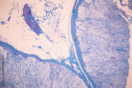
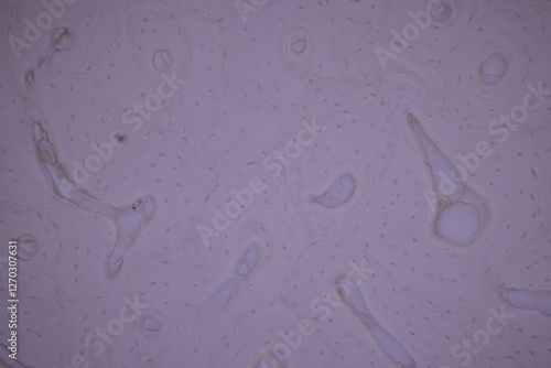
Simple squamous epithelium (mesothelium). Epithelium that lines the pleura, peritoneum, and pericardium.
Mesentery. Illustration of the 19th century. Germany. White background.
Roll of peritoneum with garlic and spices.The upper projection.Uzbek cuisine.
Crépine de porc
Baked slice of pork peritoneum with layers of fat with vegetables on a wooden table. Traditional meat snack. Copy space
Undifferentiated adenocarcinoma. Metastatic nodule
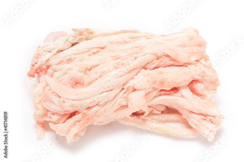
Raw pork belly with rind, peritoneum meat, spices and herbs
Lagestroemia: particolare di un fiore ancora in bocciolo
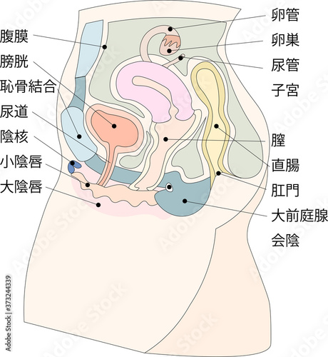
女性生殖器イラスト3日本語説明付き
dialysate cleansing home care tube fluid drain bag waste High blood cysts center manual port End Stage renal ESRD Infusion Diffusion fatty liver tap cancer B C Use pain cavity portal peritoneocentesis
Punktion
sacrum, coccyx, ilium, ischium, and pubis fuse to form the bony girdle.