Login
Sign Up
Member Area
Login
Sign Up
English
Afrikaans formal
Afrikaans informal
Arabic
Azerbaijan
Brazilian
Bulgarian
Catalan
Chinese simplified
Chinese traditional
Croatian
Czech
Danish
Dutch
English
Estonian
Finnish
French
Georgian
German
Greek
Hebrew
Hungarian
Icelandic
Indonesian
Italian
Japanese
Latvian
Lithuanian
Malaysian
Norwegian
Persian
Polish
Portuguese
Romanian
Russian
Serbian
Slovakian
Slovenian
Spanish
Swedish
Thai
Turkish
Ukrainian
Vietnamese
Toggle navigation
VideotonStock
Your Shopping Cart is empty.
Files
Stock
Photo
Video
Special offer
Collections
Packages
Lightboxes
Licenses
Royalty free
Rights managed
Creative
Editorial
Items
Most downloaded
Featured
Most popular
New
Free
Categories
Browse categories
Site info
Site info
About
Support
Privacy Policy
Privacy Policy
FAQ
Contact Us
Contact Us
Terms and Conditions
Buyer Agreement
Seller Agreement
Customers
Users
Credits
Photographers
Boby Richards
0
Boby Richards
0
Videoton1
286
Search
Keywords:
Creator ID:
Type:
Photo
Illustration
Vector
Video
Template
3d
Categories:
Animals
Buildings and Architecture
Business
Drinks
The Environment
States of Mind
Food
Graphic Resources
Hobbies and Leisure
Industry
Landscapes
Lifestyle
People
Plants and Flowers
Culture and Religion
Science
Social Issues
Sports
Technology
Transport
Travel
News
Entertainment
Sport News
Languages:
Brazilan Portuguese - Brazil
English - Canada
French - Canada
Spanish - Mexico
English - United States
English - Belgium
French - Belgium
Dutch - Belgium
Czech - Czech Republic
English - Cyprus
Danish - Danmark
German - Germany
English - Estonia
Spanish - Spain
French - France
French - Marocco
English - Greece
English - Ireland
Italian - Italia
English - Latvia
English - Lithuania
German - Luxembourg
English - Luxembourg
French - Luxembourg
English - Hungary
English - Malta
Dutch - Nederlands
Bokmal Norwegian - Norway
German - Austria
Polish - Poland
Portuguese - Portugal
English - Romania
German - Switzerland
English - Slovenia
Slovakian - Slovakia
French - Switzerland
English - Finland
Swedish- Sweden
Italian - Switzerland
English - United Kingdom
English - Bulgaria
English - Australia
Japanese - Japan
Korean - South Korea
English - New Zealand
Russian - Russia
English - Ukrain
English - Thailand
Orientation:
All
Horizontal
Vertical
square
Color:
Model property release:
All
Yes
No
Duration Video:
All
Up to 10 seconds
Up to 20 seconds
Longer than 30 seconds
Results
(0)
Site
Getty/iStock
Bigstockphoto
Pixabay
Adobe Stock
Relevance
New
Most popular
Most downloaded
Undiscovered
12
24
36
48
96
Auto paging
Menu
3D Illustration of Choroid Pigment Cells – Melanophores and Melanin-Laden Macrophages in Eye Anatomy
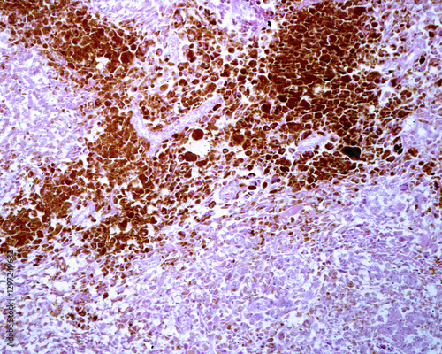
Lymph node. Melanoma metastasis
Abstract porous sphere, microscopic view.
Respiratory Epithelium Cell Structure Illustration. Ciliated, Basal, and Goblet Cells Diagram. Mucociliary Epithelium Cross Section Illustration
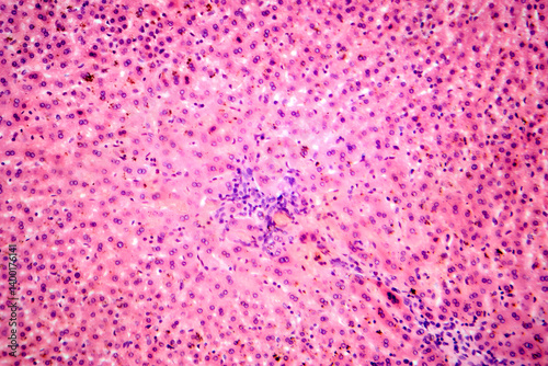
Light micrograph depicting a liver needle biopsy in haemochromatosis. This liver biopsy reveals hepatocytes with distinct brown iron deposits and highlights a fibrotic portal tract at the top.
Microscopic view of a cellular structure, showing intricate details of organelles and membranes with golden hue.
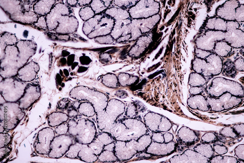
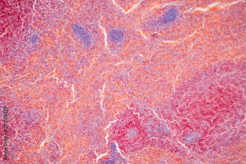
Showing Light micrograph of the Adrenal gland, Spleen human and Other human tissues under the microscope for education in the laboratory.
Intestine Cancer Cells Microscopic View
SEM micrograph of porous material structure
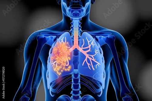
Anatomical Illustration of Lungs with Glowing Highlights Respiratory System Focus
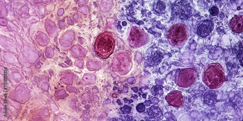
Photomicrographic image showing Metastatic squamous cell carcinoma of a lymph node, a type of skin cancer.
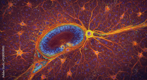
Microscopic View of Neuron Network.
Malignant cells, bronchoalveolar lavage (BAL) adenocarcinoma. Microscopic show cellular material composed of atypical epithelial cells, acute and chronic inflammatory cells.
Scanning Electron Micrograph of a Tardigrade
Abstract cellular structure, intricate network of blue and orange cells forming a mesmerizing pattern.
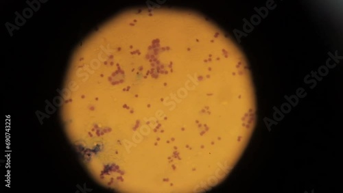
Microscopic view of Staphylococcus aureus cell under microscope.
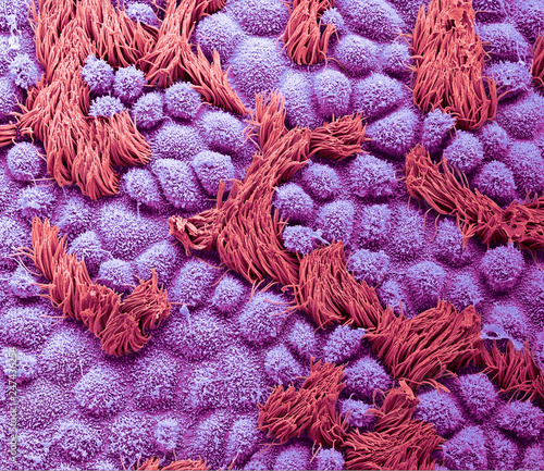
Detailed scanning electron micrograph of the hard palate lining showcasing intricate microstructures and porous textures.
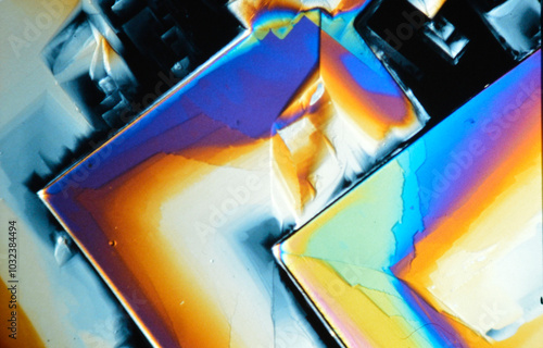
Polarized light micrograph of crystallized vitamin C
Skeletal muscle atrophy, photomicrograph showing decreased fiber size with increased spacing between them, reduced myofibrils, increased endomysial connective tissue with fatty infiltration
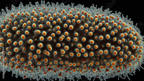
Close-up microscopic view of an organism with textured surface and orange dots
3D Illustration of Choroid Pigment Cells – Melanophores and Melanin-Laden Macrophages in Eye Anatomy
Respiratory epithelium. Cilia
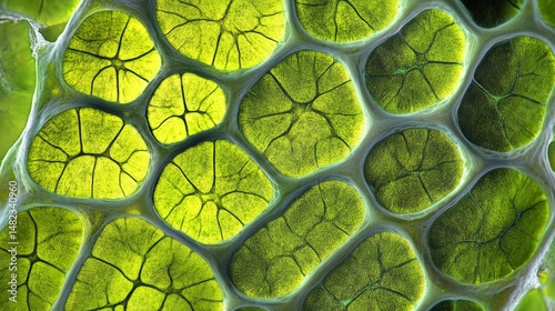
Close-up view of plant cells. AI.
cell division microscopic image png
Colored scanning electron micrograph (sem) of a tardigrade, also known as a water bear, walking on a surface
Background of human cells tissue of lung human on the microscope in pathology lab.View in microscopic of squamous cell carcinoma from human cervical cancer tissue.
Showing Light micrograph of the Adrenal gland, Spleen human and Other human tissues under the microscope for education in the laboratory.
Agarwood plant cells micrograph
Macro blue science experiment bubble texture,A close-up of a blue liquid with bubbles in a petri dish, showcasing a captivating microscopic view. ideal for concepts of science, medicine, research,
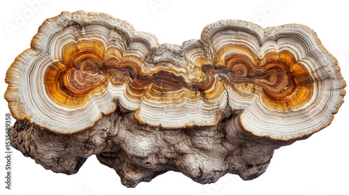
Iriartea deltoidea trunk section Transparent background, PNG file
A type of bone marrow cancer known as multiple myeloma, commonly called myeloma.
Photomicrographic image showing Metastatic squamous cell carcinoma of a lymph node, a type of skin cancer.
Light Micrograph of Blood Vessel Containing Red Blood Cells in Capillary Microcirculation
Microscopic view of cells and tissue, vibrant purple and orange clusters, detailed scientific medical background
Microscopic View of a Nerve Cell.
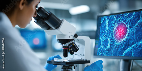
Personalized medical technology advances as scientist examines cell samples under microscope in laboratory setting for research innovation
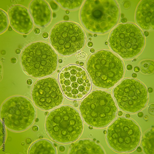
Microscopic view of the cells of a plant under the microscope.
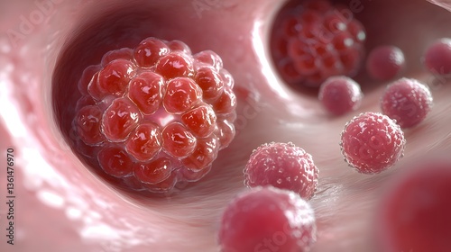
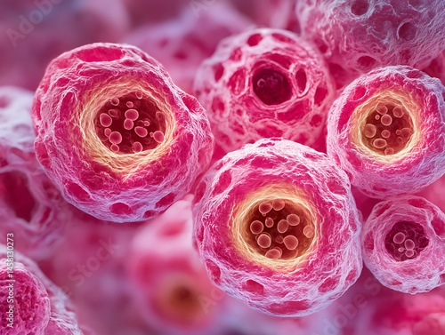
Realistic microscopy image of breast cancer cells forming a cluster with irregular shapes and high mitotic activity.
Close-Up of Glittery DNA Strand with Bright Glowing Particles on Blue Background
Skeletal muscle atrophy, photomicrograph showing decreased fiber size with increased spacing between them, reduced myofibrils, increased endomysial connective tissue with fatty infiltration
Extreme Macro Photograph of a Small Snail's Eye Stalks Highlighting Its Iridescent Features
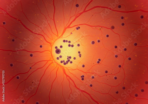
Choroid Stroma Vascular Network – Detailed Microscopic View of Blood Vessels and Pigment Cells with Melanin
Human skin. Squamous cell carcinoma
Leaf stoma under microscope during photosynthesis
Blood vessels and abundant pigment cells rich in large melanin granules observed in choroid stroma.
Abstract micrograph of a single scale from a sockeye salmon.
Fallopian tube, SEM
Backgrounds of human cells tissue of lung human under the microscope in pathology lab.View in microscopic of squamous cell carcinoma from human cervical cancer, tissue section by H and E
Showing Light micrograph of the Adrenal gland, Spleen human and Other human tissues under the microscope for education in the laboratory.
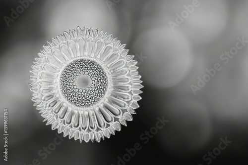
Intricate, glass-like diatom resembling a sunflower, showcasing radial symmetry and complex cellular structure.
Iriartea deltoidea trunk section Transparent background, PNG file
Willow stem, cross section, under the light microscope. Stem of Salix, also known as sallows and osiers, 8X light micrograph. Hematoxylin-eosin stained for better visualization. Isolated, over white.
Photomicrographic image showing Metastatic squamous cell carcinoma of a lymph node, a type of skin cancer.
rabbit epididymis cross section under the microscope showing spermatozoa inside its tubes - optical microscope x100 magnification
Blood sample tube and paper order request for factor VIII.
Thin skin. Epidermis
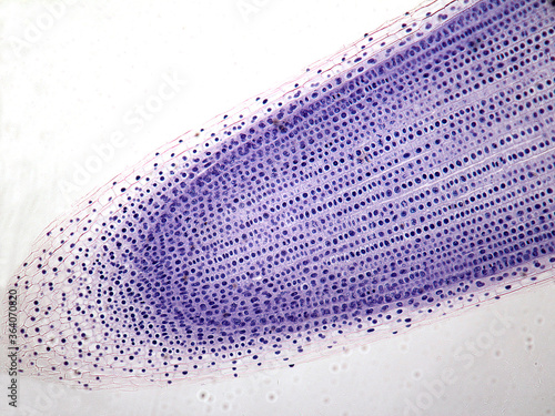
Microphotography of root of onion.
Microscopic view of Staphylococcus aureus cell under microscope.
Light micrograph showcasing human brain tissue, highlighting the intricate details of neurons and glial cells. This light micrograph captures the essential characteristics of brain tissue.
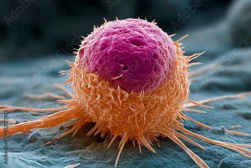
color-enhanced SEM style cancer cell, realistic texture, subtle shadows, not oversaturated
Coccidia in liver, photomicrograph showing intracellular parasitic protozoa causing hepatic coccidiosis with inflammation and hepatocellular damage.
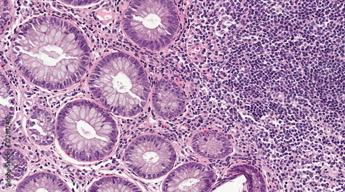
Mildly inflamed colonic mucosa - microscopic image 20x
Close-up Light Micrograph Showing Human Brain Neurons and Glial Cells in Detail
Respiratory epithelium. Cilia
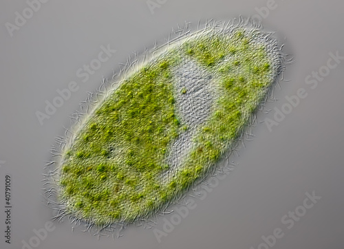
Paramecium bursaria - grünes Pantoffeltierchen - DIC
Pine Wood micrograph
Crystals of a common painkiller, Aspirin
Backgrounds of human cells tissue of lung human under the microscope in pathology lab.View in microscopic of squamous cell carcinoma from human cervical cancer, tissue section by H and E
Showing Light micrograph of the Adrenal gland, Spleen human and Other human tissues under the microscope for education in the laboratory.
Microscopic view of cells with abstract patterns in purple hues.
Iriartea deltoidea trunk section Transparent background, PNG file
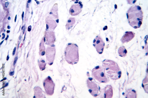
Bone marrow's largest cell, the megakaryocyte, plays a crucial role in blood platelet production.
Photomicrographic image showing Metastatic squamous cell carcinoma of a lymph node, a type of skin cancer.
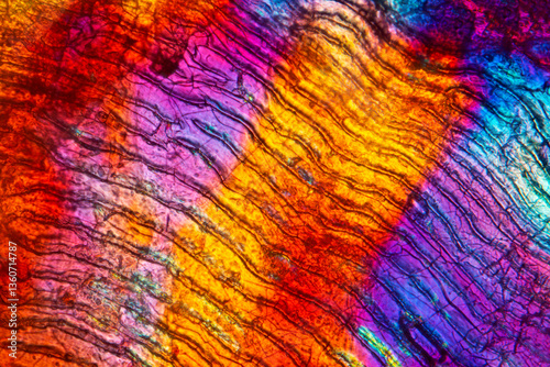
Digitally manipulated, abstract micrograph of olivine pyroxenite with polariztion.
Blood sample tube and paper order request for factor VII.
Licmophora sp. algae, marine and freshwater diatom under microscopic view. Genus of benthic, photosynthetic and epiphyte diatom
Microphotography of root mitosis of an onion.
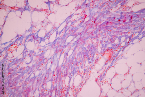
Human fat body tissue under microscope view for physiology education
Polarised light micrograph of a salt crystal, sodium chloride
Mesmerizing Blue Eyes of Woman Wearing Intricate Veil in Ethnic Attire
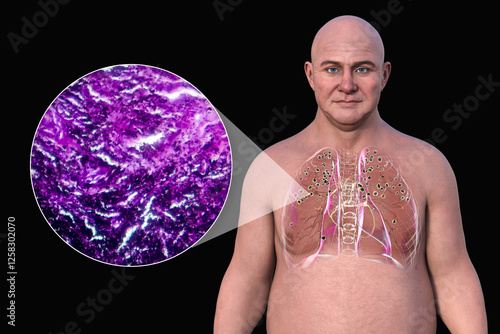
Man with silicosis, 3D illustration and light micrograph
Radiant Mineral Crystal Formation with Faceted Surfaces and Vivid Color Play
Choroid Stroma Vascular Network – Detailed Microscopic View of Blood Vessels and Pigment Cells with Melanin
Human endometrial glands. Early secretory phase
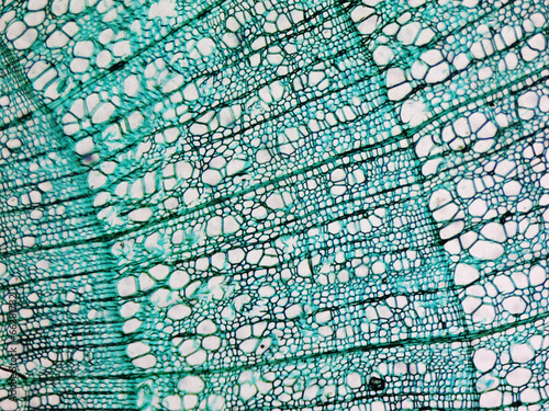
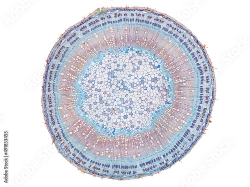
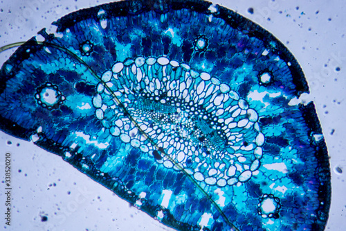
Micrograph of plant cells in a woody dicot stem showing vascular tissue structure and cell wall details under a microscope for botanical and scientific education use
Cotton stem micrograph transparent PNG
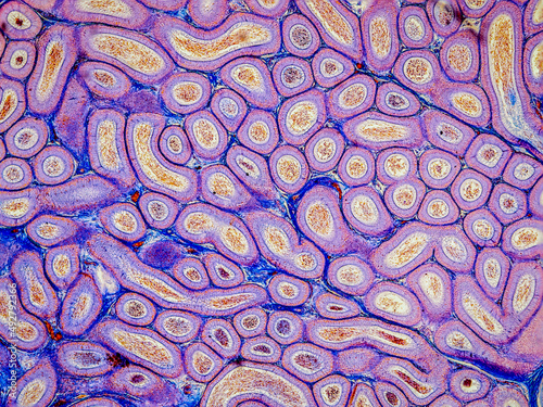
Pine leaf, cross section, 20X light micrograph. Pine tree needle under the microscope. Epidermis cells, cuticle, mesophyll, resin canals, central oval with vascular cylinder surrounded by endodermis.
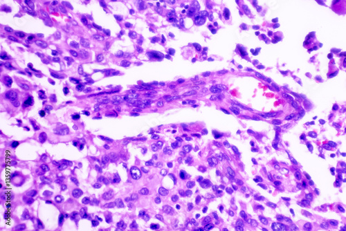
Light micrograph depicting a smear with cellular elements. Cancer. Inflammatory smear exhibiting relevant changes.
Showing Light micrograph of the Adrenal gland, Spleen human and Other human tissues under the microscope for education in the laboratory.
cross section cut slice of plant stem under the microscope – microscopic view of plant cells for botanic education
Iriartea deltoidea trunk section Transparent background, PNG file
biopsy. biopsy results under a microscope. Human cancer cells under a microscope. human cells taken as a result of a biopsy for study in a laboratory using professional equipment. Anatomical theme.
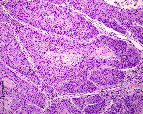
Microscopic image of Trephine Biopsy showing Acute Leukemia, Bone marrow biopsy
Micrograph of a papillose moss leaf, with polarization at 400x.
Medical themes: Microscopic image of breast cancer with capillary arrangement
Microscopic image of pine leaf cross-section
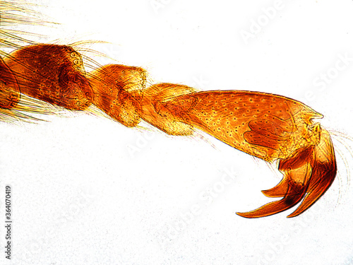
Microphotography of leg of bee. Apis mellifera.
Blood sample tube and paper order request for PTT.
rotifera of the family Habrotrochidae under the microscope - light microscope x400 magnification
Chronic lymphocytic leukemia is the most prevalent type of chronic leukemia among adults. Blood smears reveal elevated levels of mature lymphocytes and smudge cells, which are remnants of unusually