Login
Sign Up
Member Area
Login
Sign Up
English
Afrikaans formal
Afrikaans informal
Arabic
Azerbaijan
Brazilian
Bulgarian
Catalan
Chinese simplified
Chinese traditional
Croatian
Czech
Danish
Dutch
English
Estonian
Finnish
French
Georgian
German
Greek
Hebrew
Hungarian
Icelandic
Indonesian
Italian
Japanese
Latvian
Lithuanian
Malaysian
Norwegian
Persian
Polish
Portuguese
Romanian
Russian
Serbian
Slovakian
Slovenian
Spanish
Swedish
Thai
Turkish
Ukrainian
Vietnamese
Toggle navigation
VideotonStock
Your Shopping Cart is empty.
Files
Stock
Photo
Video
Special offer
Collections
Packages
Lightboxes
Licenses
Royalty free
Rights managed
Creative
Editorial
Items
Most downloaded
Featured
Most popular
New
Free
Categories
Browse categories
Site info
Site info
About
Support
Privacy Policy
Privacy Policy
FAQ
Contact Us
Contact Us
Terms and Conditions
Buyer Agreement
Seller Agreement
Customers
Users
Credits
Photographers
Boby Richards
0
Boby Richards
0
Videoton1
286
Search
Keywords:
Creator ID:
Type:
Photo
Illustration
Vector
Video
Template
3d
Categories:
Animals
Buildings and Architecture
Business
Drinks
The Environment
States of Mind
Food
Graphic Resources
Hobbies and Leisure
Industry
Landscapes
Lifestyle
People
Plants and Flowers
Culture and Religion
Science
Social Issues
Sports
Technology
Transport
Travel
News
Entertainment
Sport News
Languages:
Brazilan Portuguese - Brazil
English - Canada
French - Canada
Spanish - Mexico
English - United States
English - Belgium
French - Belgium
Dutch - Belgium
Czech - Czech Republic
English - Cyprus
Danish - Danmark
German - Germany
English - Estonia
Spanish - Spain
French - France
French - Marocco
English - Greece
English - Ireland
Italian - Italia
English - Latvia
English - Lithuania
German - Luxembourg
English - Luxembourg
French - Luxembourg
English - Hungary
English - Malta
Dutch - Nederlands
Bokmal Norwegian - Norway
German - Austria
Polish - Poland
Portuguese - Portugal
English - Romania
German - Switzerland
English - Slovenia
Slovakian - Slovakia
French - Switzerland
English - Finland
Swedish- Sweden
Italian - Switzerland
English - United Kingdom
English - Bulgaria
English - Australia
Japanese - Japan
Korean - South Korea
English - New Zealand
Russian - Russia
English - Ukrain
English - Thailand
Orientation:
All
Horizontal
Vertical
square
Color:
Model property release:
All
Yes
No
Duration Video:
All
Up to 10 seconds
Up to 20 seconds
Longer than 30 seconds
Results
(0)
Site
Getty/iStock
Bigstockphoto
Pixabay
Adobe Stock
Relevance
New
Most popular
Most downloaded
Undiscovered
12
24
36
48
96
Auto paging
Menu
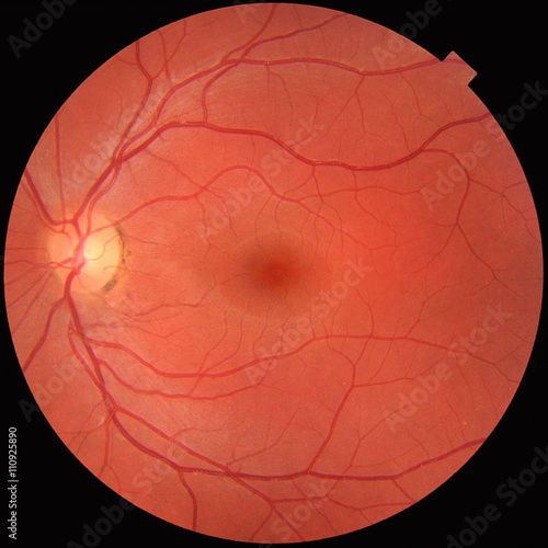
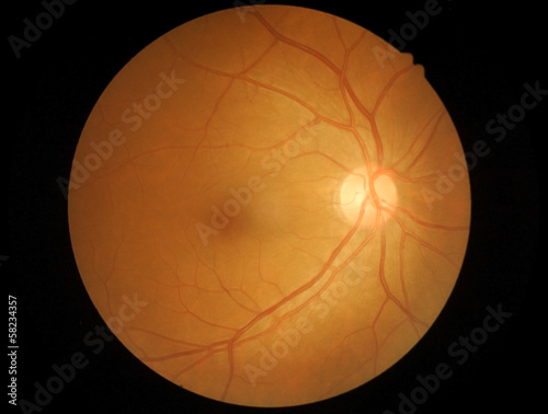
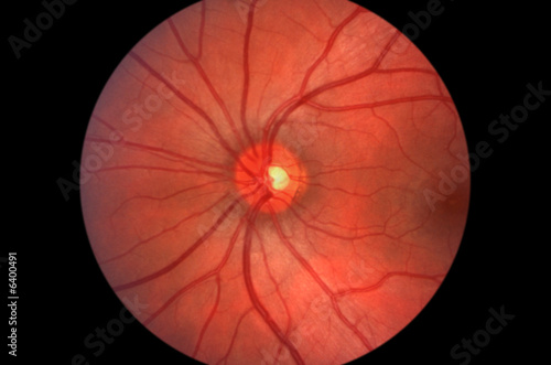
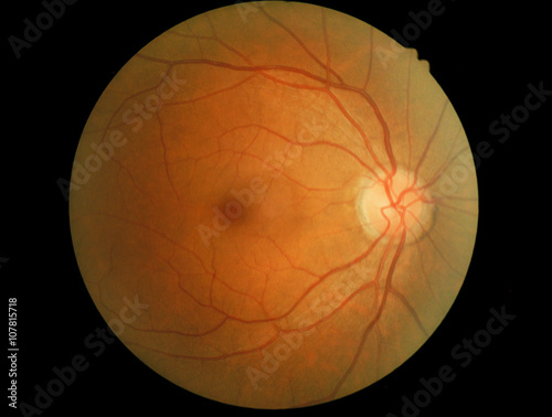
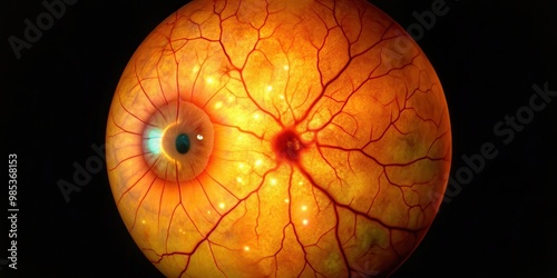
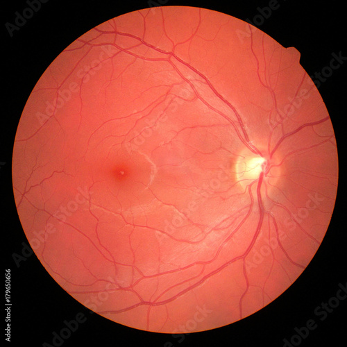
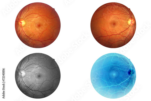
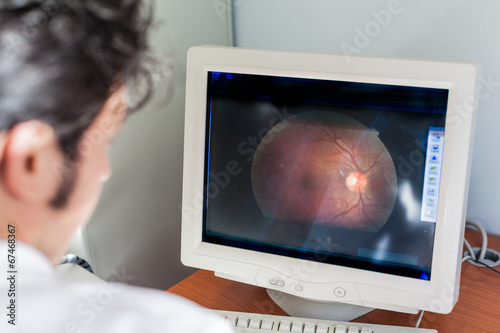
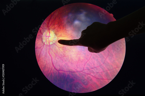
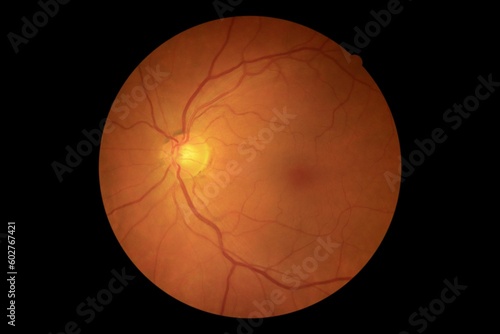
Fundus photograph of a normal left eye. Macula in center and optic disk where blood vessels converge with pigmentation on perimeter
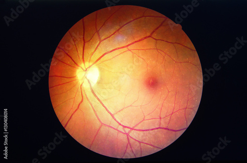
diabetes retinopathy.
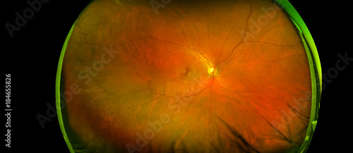
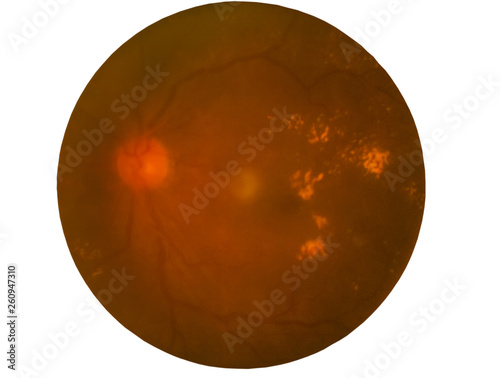
Photo Madical Retina Abnormal isolated on black background.Retina of diabetes diabates retinopathy.
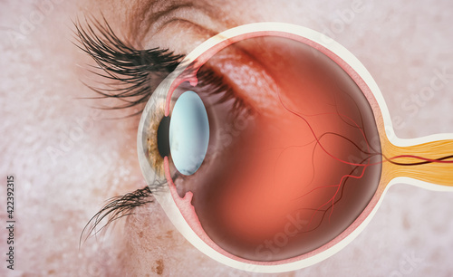
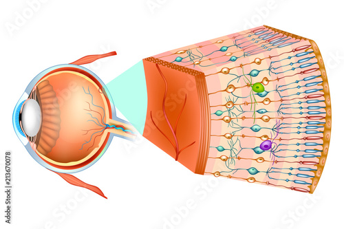
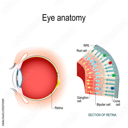
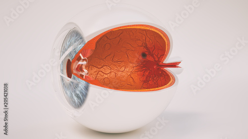
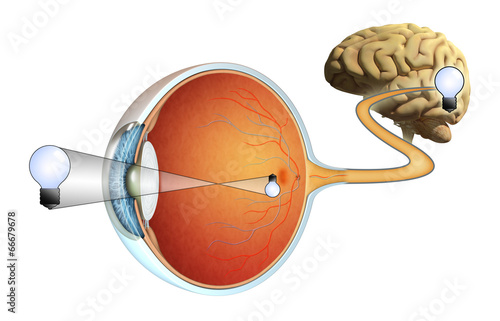
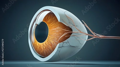
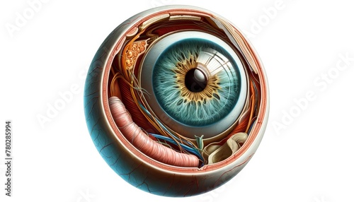
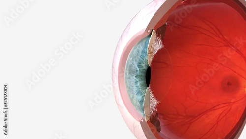
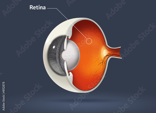
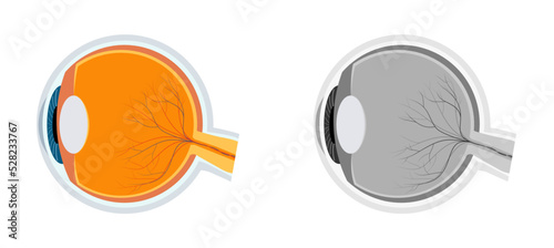
Structure of human eye. In side view.
Medical photo retina and optic nerve
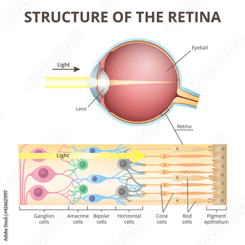
Structure of the human eye and organization of the retina. Optic part of retina.
A Comprehensive Overview of Glaucoma and Its Effects on Sight
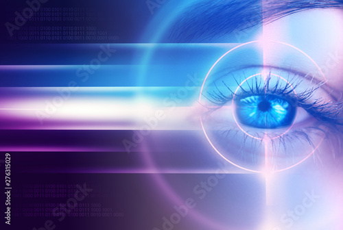
retina scan concept
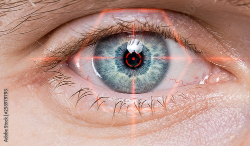
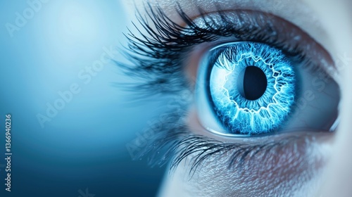
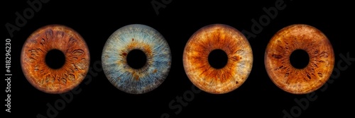
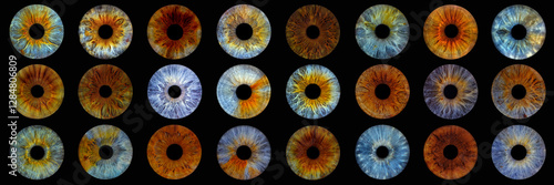
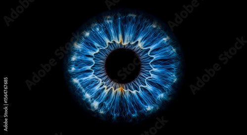
eye iris
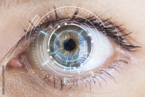
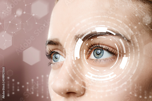
Multilayer security using fingerprint and retina scan to prevent fraudulent transactions
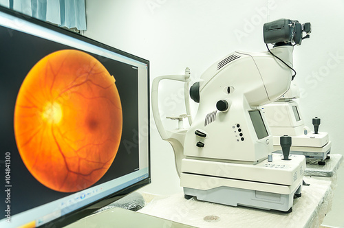
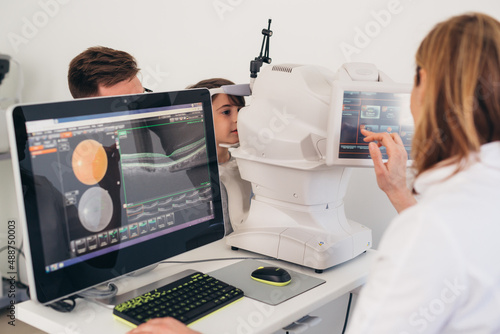
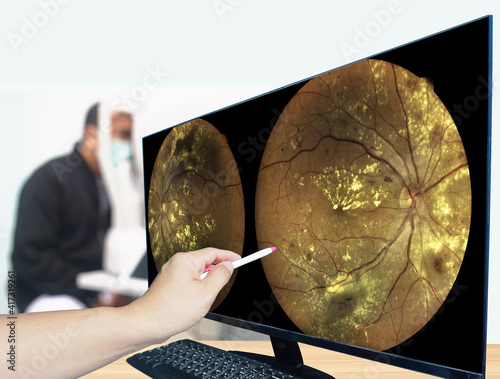
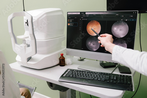
fundus camera use for examination eye in hospital
Research and scanning eye, close-up photos, retinal diagnostics in ophthalmology
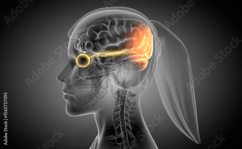
Woman with optic nerve and link to stimulated brain region - X ray style 3D illustration with dark gray background
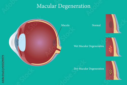
Close-up view of retina scan revealing yellow deposits associated with macular degeneration
Retina - Optic Nerve - Human Eye
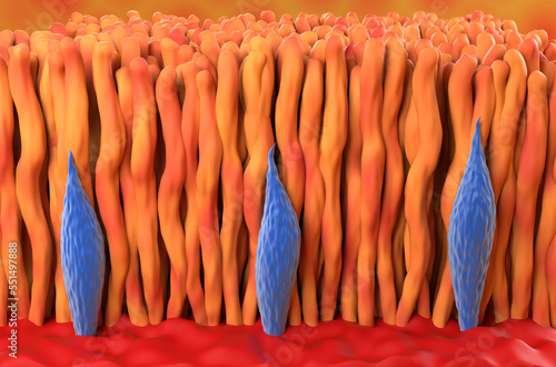
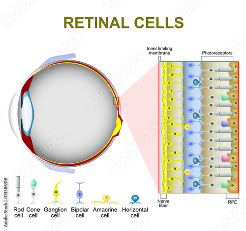
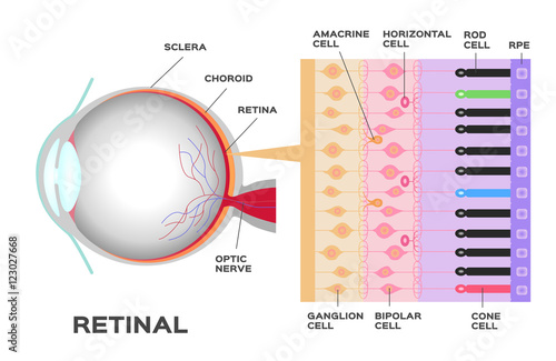
Eye anatomy. Rod cells and cone cells.
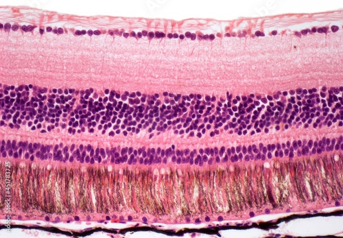
Retina, LM
Abstract visualization of an eye with yellow blue red retina and iris as conceptual background for science and research or futuristic wallpaper with copy space for text
eye's retinal angle image with macula, vessels and optic disc isolated view on a black bacground. made by ultra wide fundus camera
Human eye anatomy, retina, optic disc artery and vein etc. takin
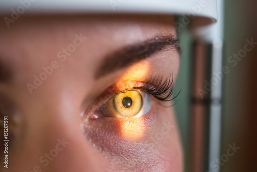
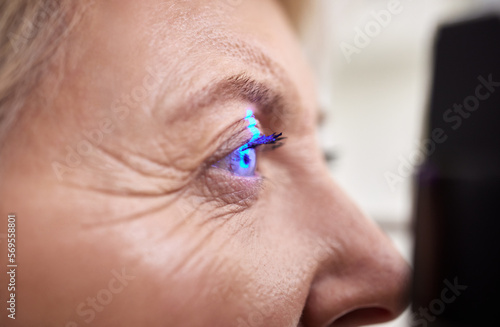
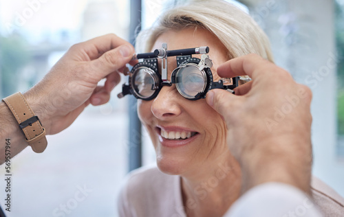
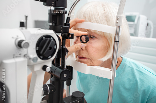
Laser, vision or senior customer in eye exam for eyesight at optometrist office in assessment or consultation. Face of mature woman testing or checking vision to help iris or retina visual health
child having eye examination.digital retina scan
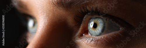
Amazing female green colored eyes in low light technique
Human eye anatomy, retina, optic disc artery and vein etc.
Ophthalmic image detailing the retina and optic nerve inside a healthy human eye. Medicine concept
Menschliches Auge im Querschnitt - sehr detailreich
Anatomy of normal eye and Diabetic retinopathy in people who have diabetes. Illustration about health and eyesight.
Resolution An eye looking at a retina display screen with extreme clarity and precision.
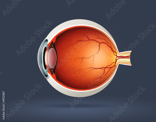
Human eye cross section
Close-up of a biometric retina scanner
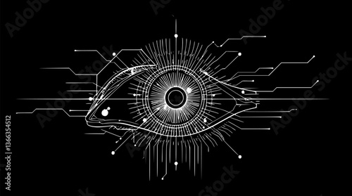
Outline of an AI-powered eye minimal biometric scan technology vector art.
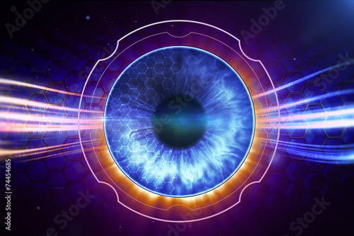
Retina Scanner
Optical coherence tomography OCT scan to create pictures of the back of eye. Examination of eyes of patient using optical coherence tomograph. Ophthalmologist is scanning cornea of girl patient.
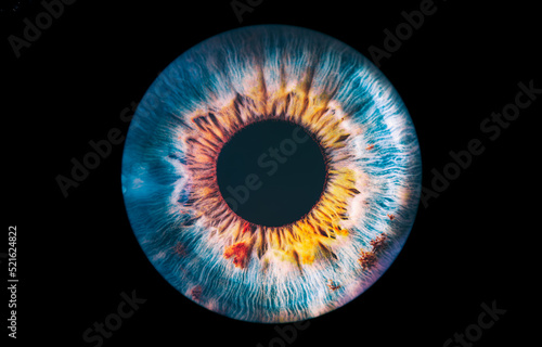
Close up of eye iris on black background, macro, photography
Vision
This text provides an informative look at macular degeneration, featuring a model eye designed for educational purposes
of pigment deposits in the retina caused by retinitis pigmentosa disease, genetic, eye disease
Eye viewing digital information
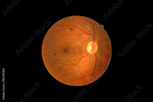
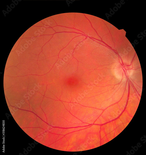
left eye's retinal image with macula, vessels and optic disc isolated view on a black bacground
Eye Exam: Close-up of a medical professional examining a patient's blue eye with specialized equipment.
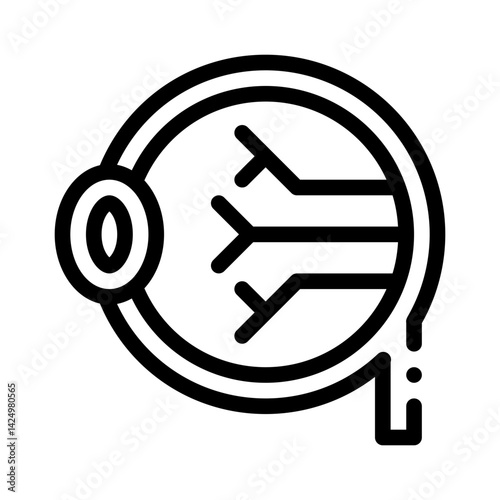
retina line icon
older woman has eye examination. digital retina scanner
Detailed cross section of the human eye anatomy highlighting the retina cornea and optic nerve structures This medical diagram showcases the intricate anatomical features of the eye
Human eye anatomy, retina, optic disc artery and vein etc.
Retina cone and rod in the human eye – isometric view 3d illustration
3d illustration of the human eye anatomy
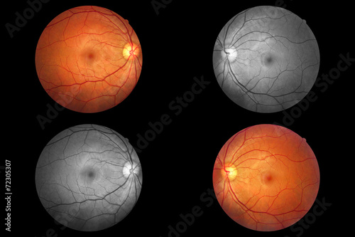
Fundus photography Madical Retina both eye Abnormal isolated on white background.Retina of diabetes check up medical healthcare and technology concept.
Amazing female green colored eyes in low light technique
Extreme close up human eye. Human eye iris contracting. Extreme close up.
Close up of eye iris on black background, macro, photography. Colorful human eye irises.
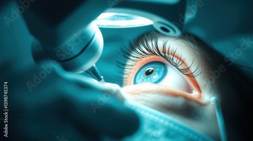
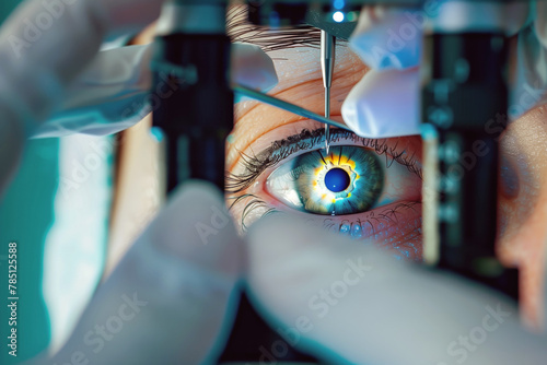
Close-up of a patient undergoing laser eye surgery
Retina
Examination of eyes of patient using optical coherence tomograph. Ophthalmologist is scanning cornea of girl patient. Optical coherence tomography OCT scan to create pictures of the back of eye.
modern technologies of scanning of the retina of the eye
Photoreceptor cells in the retina of the eye
Research and scanning eye, close-up photos, retinal diagnostics in ophthalmology
Eye exam, vision or laser test for an old woman with a machine at optometry consultation for retina problem. Senior, patient or client with medical health insurance checking eyesight at optician
Doctor, face or black woman in eye exam assessment consultation for eyesight at optometrist office. Zoom of optician helping a mature customer testing or checking vision, iris or retina visual health
A detailed illustration of a human eye, dissected to show the iris, lens, and retina in an artistic style.
Age-related macular degeneration.Eye vision disorder on green background.
Hands, eye scan and screen in optometry for doctor, consultation and examination for vision health. Person, retina and medical advice on technology in clinic for optical wellness, therapy or analysis
High-tech iris scanning process, abstract data patterns, deep blue background, iris scan, biometric security
eyeball in section, structure of the retina, close-up
Optical Coherence Tomography (OCT) image of eye in the patient. Ophthalmic test - OCT optical coherence tomography measurement. SLO Scan view of the macula in retina with vessels.
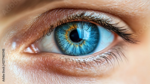
Captivating Iris: A Macro View of Stunning Blue Eye Detail - Vision, Anatomy, and the Beauty of Human Eyes
Optometrist demonstrating eye anatomy during patient consultation in clinical setting. Vision treatment and correction, eye care
The cornea is the transparent front part of the eye that covers the iris, pupil, and anterior chamber
Ophthalmology care, eyesight or vision. Asian young woman, girl hand holding and putting, wearing soft, daily disposable contact lens on her face with finger, looking into eye. Medical and health care
Biometric security system scanning retina, high-tech background, cold light, cyber security concept, photorealistic
Diagnosis eye diabetes.
Close up woman eyes. Macro classic volume long eyelash extensions, lamination, correction. Natural beauty or laser vision correction. Skin care, beauty, makeup concept.
Detailed assessment of eye health and vision. Management of cataracts glaucoma
Retinal scan analysis for vision health, optic nerve evaluation, and early detection at ophthalmology. Ophthalmologist examines retina images on screen, diagnosing glaucoma and macular degeneration.
Retina
Eye of the female on a dark abstract background, neon holograms, retina scanner
eye's retinal angle image with macula, vessels and optic disc isolated view on a black bacground. made by ultra wide fundus camera
Hands, help or woman in eye exam or vision test for eyesight by doctor, optometrist or ophthalmologist. Optician helping a happy customer to see or check glaucoma or retina health in a consultation
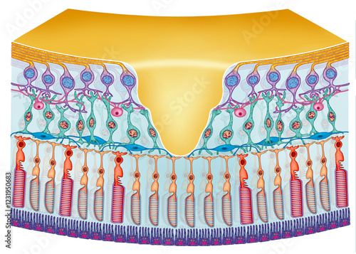
Schematic illustration of a segment of the retina, showing the light-sensitive photoreceptor cells.
Cataract and Retina Concept ,Close up of eye with detailed iris and pupil, showcasing eye movement during retina scan
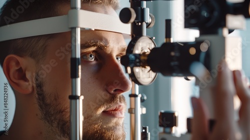
Optometrist uses laser machine to examine man's eye for retina disease, ensuring eyesight and medical insurance coverage.
Close-up of eye surgery with medical equipment in operation.
Retina Scan Biometric Technology Backdrop – Detailed image focusing on retina scanning technology.
Diagram of the retina of the eye
eye infographic: Photoreceptor in the retina of the eye. Structure and function rod and cone cells. Vector scheme
Close-up grayscale human eye
Senior woman eyesight test with binocular slit-lamp. Checking retina of a female eye close-up. Ophthalmology clinic
Human Eye Anatomy Vector Image
Abstract xray art of an Eye
Netzhaut des menschlichen Auges.
biometric hi tech security retina scan
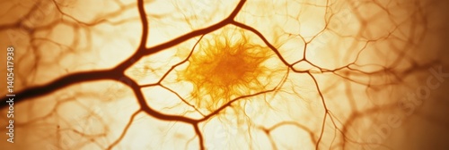
macrophotography of a human retina
Detailed Eye Anatomy Illustration Showing Internal Structures and Optical Components
Function of retina of human eye
Laser Eye Vision Correction Procedure With A Colored Dark-Skinned Lady's Eye and Laser Beam Close View Background
Retina of diabetes , diabates retinopathy,photo Medical Retina Abnormal (cataract) isolated on white background.Saved with clipping path
Colorful abstract iris 3d illustration
Young woman with iris scanning, closeup
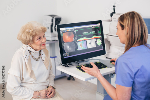
Glaucoma, Cataract, Retinal Disease Male ophthalmologist explains eye disease using an eye model to an Asian female patient at the examination table in a hospital examination room.
Close up of human retina revealing intricate neural and blood vessels, pulsing with vibrant colors and macro details.
Detailed assessment of eye health and vision. Management of cataracts glaucoma